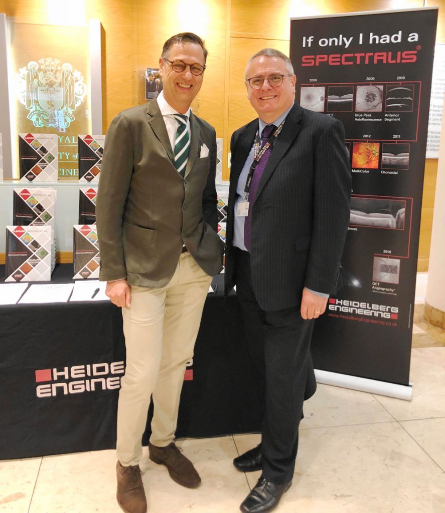
Hemel Hempstead, UK – Insight into the most advanced glaucoma imaging – from world-leading experts – was the focus at the Royal Society of Medicine last week when the new Heidelberg Engineering Glaucoma Imaging Atlas was launched.
Designed to ensure eye health professionals take full advantage of the diagnostic and monitoring capabilities of OCT technology, Heidelberg Engineering launched the reference which includes contributions from 29 internationally respected clinicians.
“The diagnostic imaging guide for glaucoma assessment and management features contributors from five countries and 30 detailed patient case studies. It is the cornerstone of Heidelberg Engineering Academy’s educational programme,” said Ali Tafreshi, Heidelberg Engineering’s Director of Clinical Research in Germany.
“Our longstanding commitment to high-quality diagnostic imaging provides a global standard in eye care practices and academic institutions. The reference highlights the power the clinician gains from looking at high-quality images to take full advantage of our technology and enhance diagnostic capability. The Atlas is designed to be used as a teaching tool to further educate the eye care community on the implementation of OCT technology into the glaucoma clinic, as an aid to diagnosis and management, enabling effective, individualized patient care,” he said.
Presentations of case studies were made by Mr Tafreshi and Christopher Mody, UK Director of Clinical Affairs, and Professor Christian Mardin, Senior Consultant Ophthalmologist at the University of Erlangen-Nuremberg, Germany.
Professor Mardin spoke of the socioeconomic importance of early diagnosis and treatment of glaucoma as late stage treatment and care presents a “huge economic burden” –
“The financial burden of glaucoma increases with disease severity in terms of treatment, lost ability to work, drive and the implications to mental health.”
He presented a number of case studies from his clinic and the Atlas to show how neuroretinal rim, ganglion cell and retinal nerve fibre layer measurements combined with accurate progression analysis aid the clinician in making a confident glaucoma diagnosis –
“The Atlas should motivate full use of the latest scanning technology and how modern glaucoma diagnosis benefits from the use of OCT imaging in conjunction with traditional disc photography and visual field assessment,” he said.
Contact Academy-UK@HeidelbergEngineering.com to find out how to get a Glaucoma Imaging Atlas.


