Scientific Timeline
An overview of our contributions to ophthalmic science
Navigate this scientific timeline to find out more about how Heidelberg Engineering has been researching, cooperating and continuously optimizing ophthalmic imaging and healthcare IT technologies over several decades.
Every step of the way, the goal of our many collaborations with scientists, clinicians and industry partners has been − and continues to be − to develop clinically relevant, innovative products to empower clinicians to improve patient care.
| ← | 1984 | 1988 | 1989 | 1990 | 1991 | 1994 | 1996 | 1997 | 1999 | 2000 | 2001 | 2002 | 2004 | 2005 | 2006 | 2007 | 2008 | 2010 | 2012 | 2013 | 2014 | 2015 | 2016 | 2017 | 2018 | 2019 | 2020 | → |
1984
First research prototype of adaptive optics (Heidelberg Instruments)
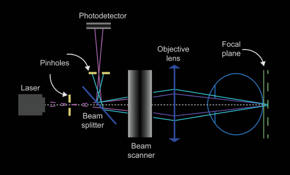
First commercial prototype cSLO (Heidelberg Instruments)
Zinser, Bille
⇩
 |
1988First Hartmann-Shack Wavefront Aberrometry sensor including Zernike Polynomials |
⇩
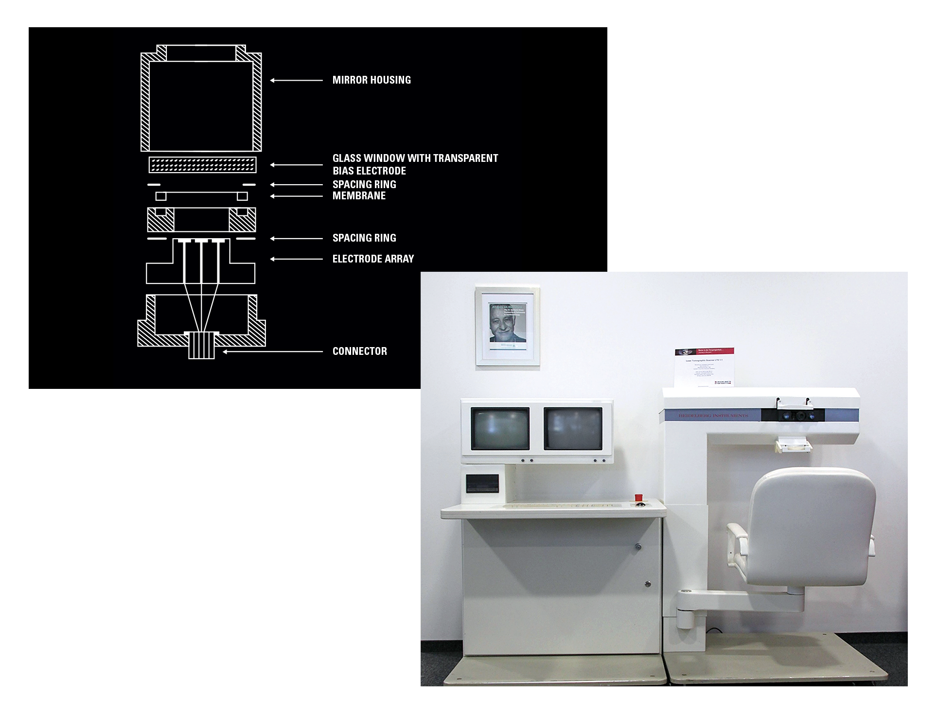
1989Aberration corrected retinal imaging First Commercial cSLO: Laser Tomographic Scanner (LTS, Heidelberg Instruments) |
 |
⇩
1990
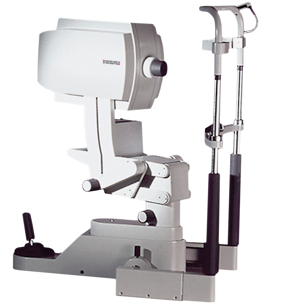
![]()
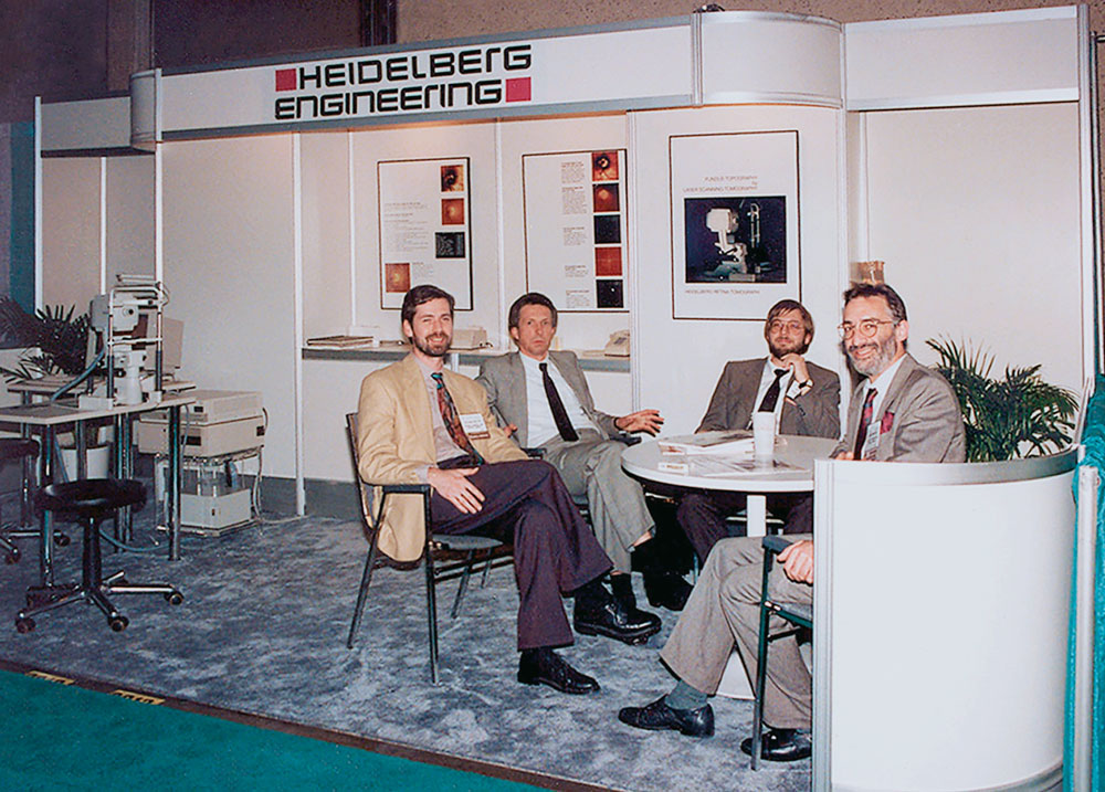
Heidelberg Engineering was formed
Zinser, Schoess
⇩
1991
First AAO with Heidelberg Engineering
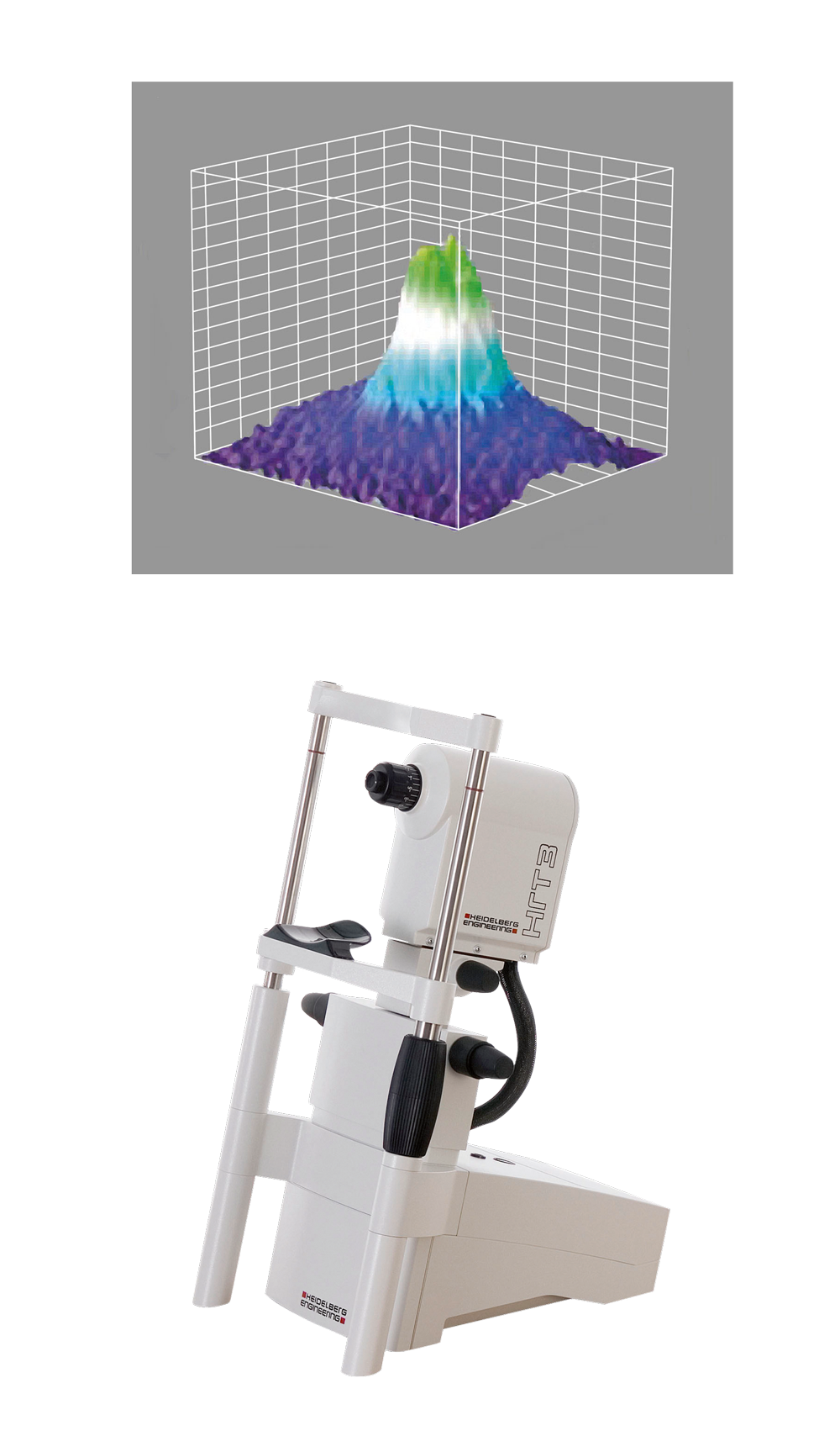
First commercial compact cSLO with objective measurement of ONH with automatic image alignment for correction of eye motion (HRT)
⇩
 |
1994First research prototype of adaptive optics (Heidelberg Instruments) |
⇩
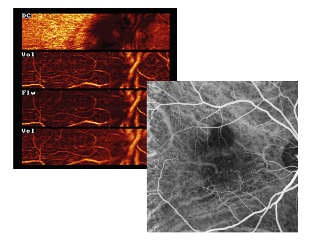
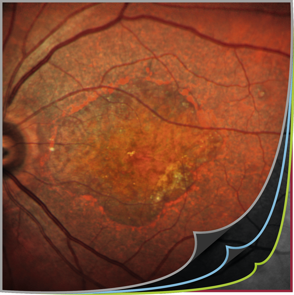
1996First Doppler Flowmetry (HRF) First ICGA study with HRA |
 |
⇩
1997
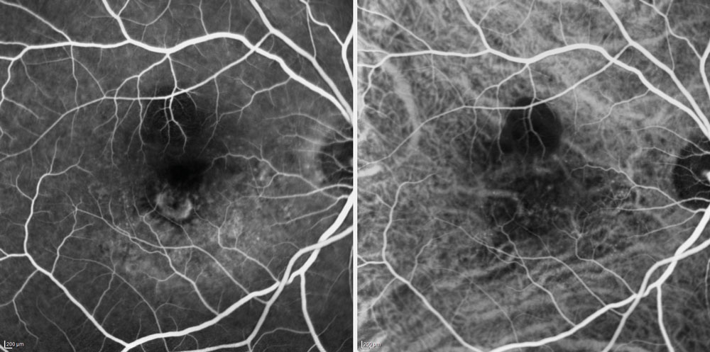
First simultaneous FA / ICGA video technology
Holz, et al.
⇩
1998
Image montage using image averaging technology on HRA
Noise reduction technology by image averaging on HRA
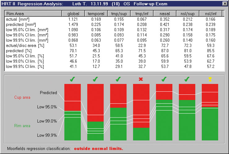
Moorfields Regression Analysis (MRA)
Wollstein, Garway-Heath, et al.
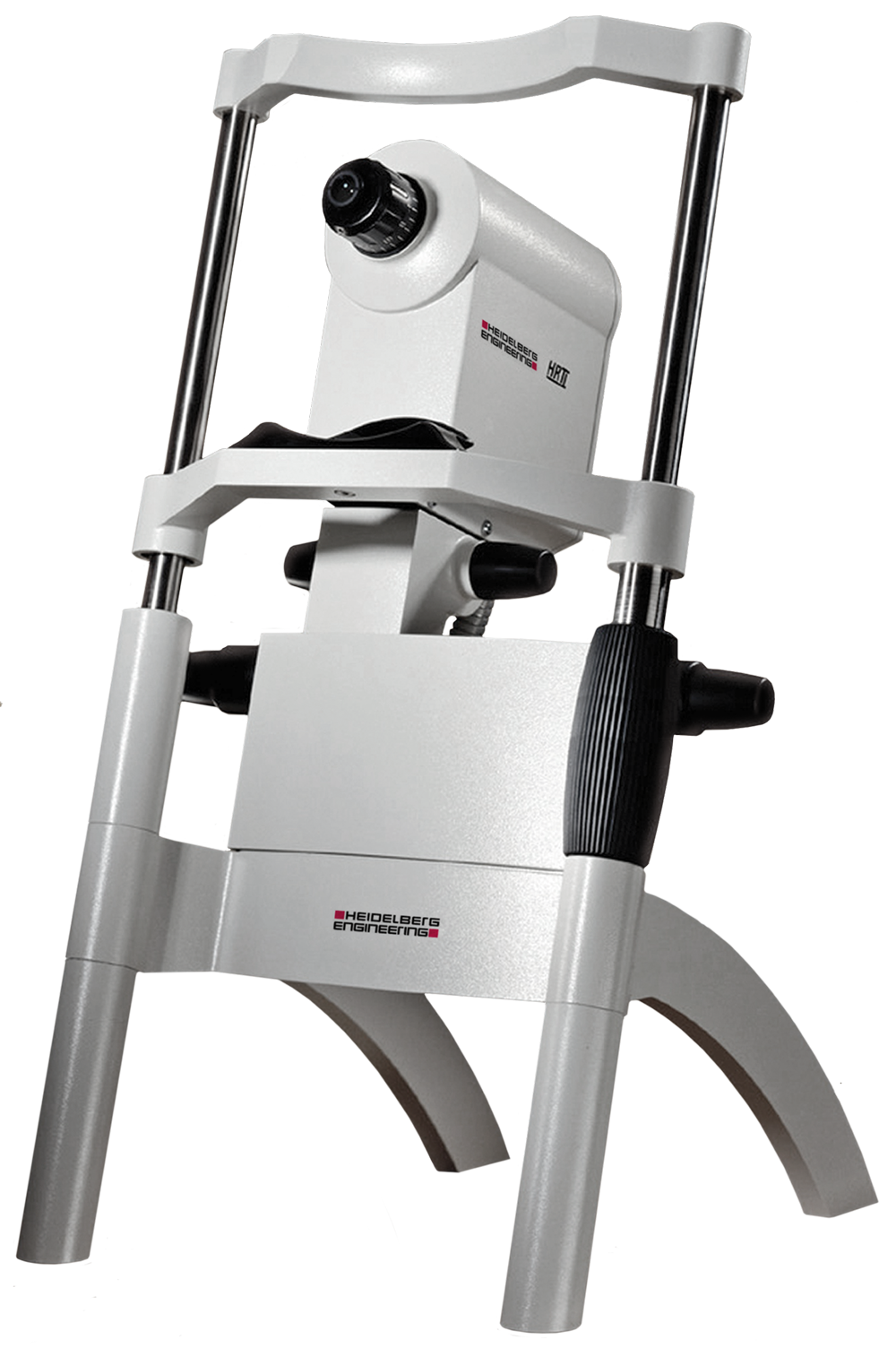
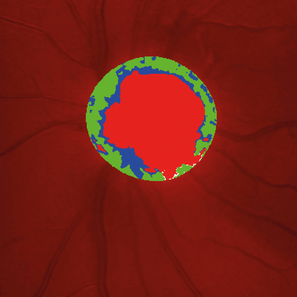
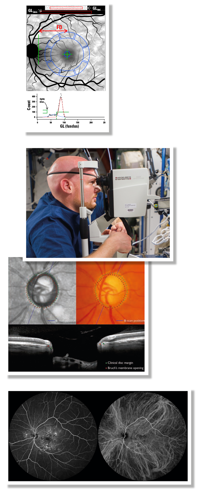
First compact clinical system for objective measurement of the ONH – became gold standard for progression analysis (HRT II)
⇩
1999
First Oral FA with HRA
Garcia, et al.
⇩
2000
Glaucoma Probability Score with HRT II
First machine learning classification algorithm (AI)
Swindale, Stjepanovic, et al.
Topographic Change Analysis (TCA)
Chauhan, et al.
⇩
 |
2001Blue Fundus Autofluorescence using HRA2 prototype |
⇩
2002
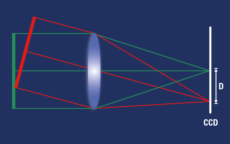
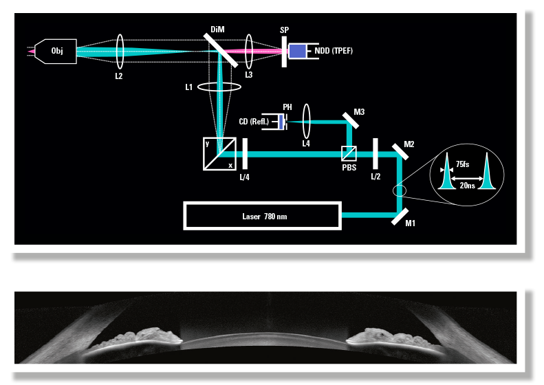
Lab prototype of compact cSLO system for two-photon imaging
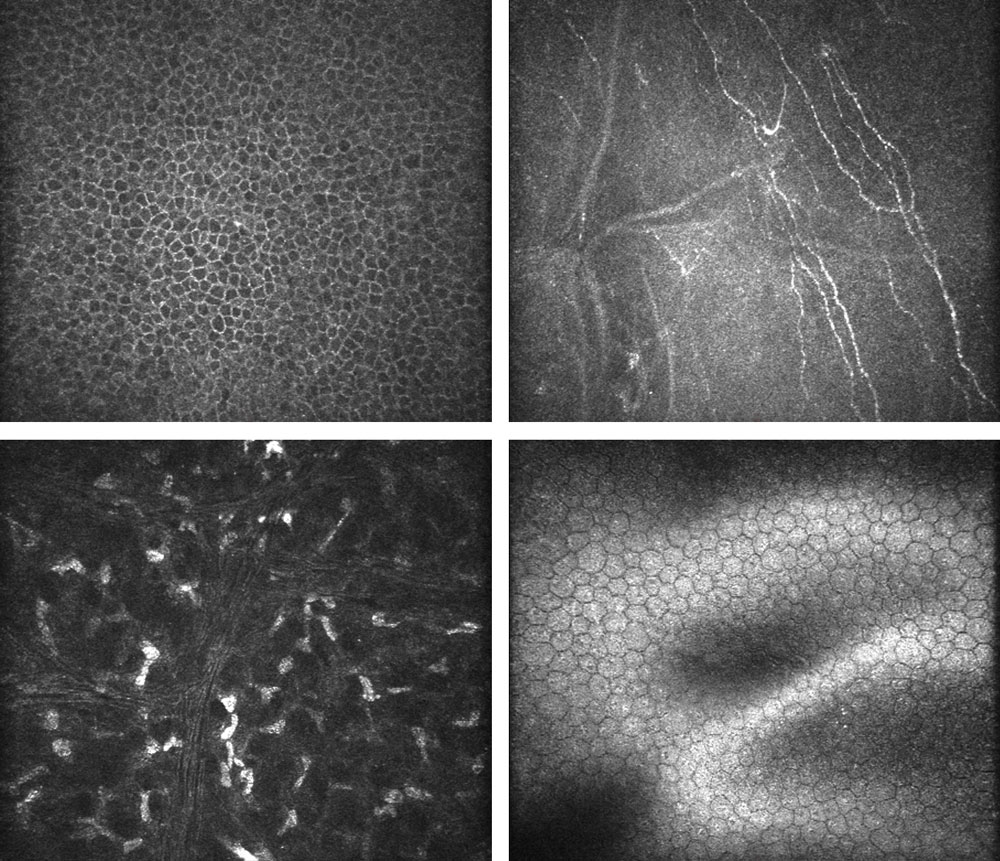
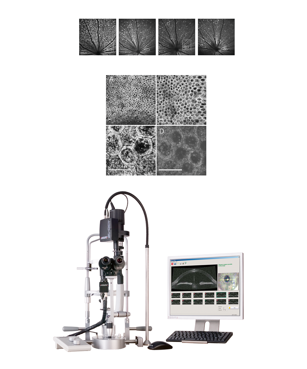
First cSLO in vivo corneal microscopy (HRTII RCM prototype)
Guthoff, Stave, Stachs, et al.
⇩
2004In vivo measurement of time resolved autofluorescence of the human eye First commercial multi-modal cSLO with real-time image averaging, noise reduction and image composites, AF and simultaneous FA / ICGA video (HRA2) |
 |
 |
⇩
 |
2005First MPOD study, using HRA First HRT3 replaces HRT II with AI (GPS), and TCA |
⇩

2006
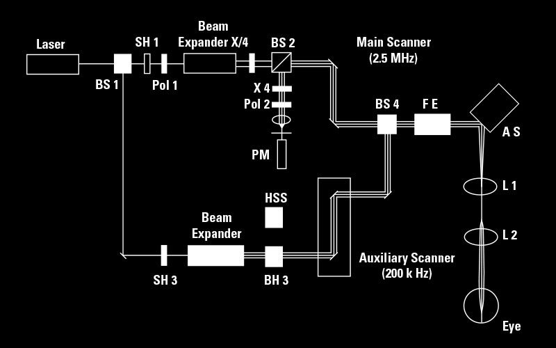
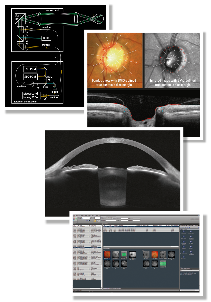
First SPECTRALIS HRA+OCT with simultaneous cSLO and OCT with real-time TruTrack eye tracking with dynamic visualization (colocalization of retina and OCT image)
First lab two-photon excited fluorescence imaging in vitro human eyes
Bille, Holz, Schneider, et al.
⇩
2007
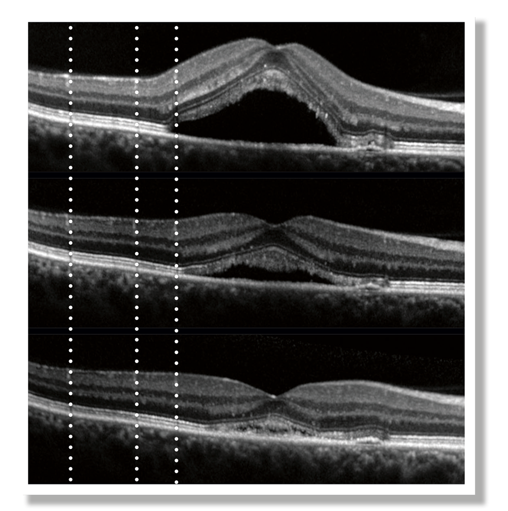
First AutoRescan using eye tracking on SPECTRALIS OCT
⇩
2008
First MultiLine study in rodents
Leung, Weinreb, et al.
First prototype for ex vivo two-photon fluorescence imaging of the human eye Slit-Lamp OCT
⇩
2010
First two-photon fluorescence imaging on in-vivo rabbit eye
Jester, La Schiazza, et al.
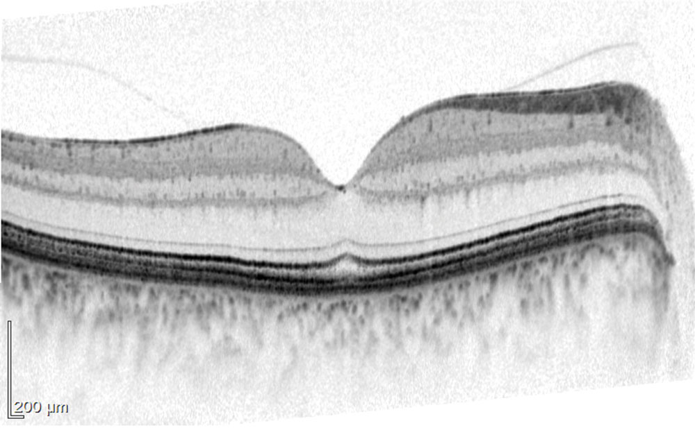
First anterior segment imaging with SPECTRALIS OCT
⇩
2012
Blue/Green/IR laser cSLO imaging (MultiColor)
⇩
2013Quantitative autofluorescence and macular pigment optical density (MPOD) cSLO NASA Ocular Health Study with SPECTRALIS OCT on ISS Prototype software for BMO-MRW with APS study Ultra-widefield FA / ICGA cSLO |
 |
⇩
2014
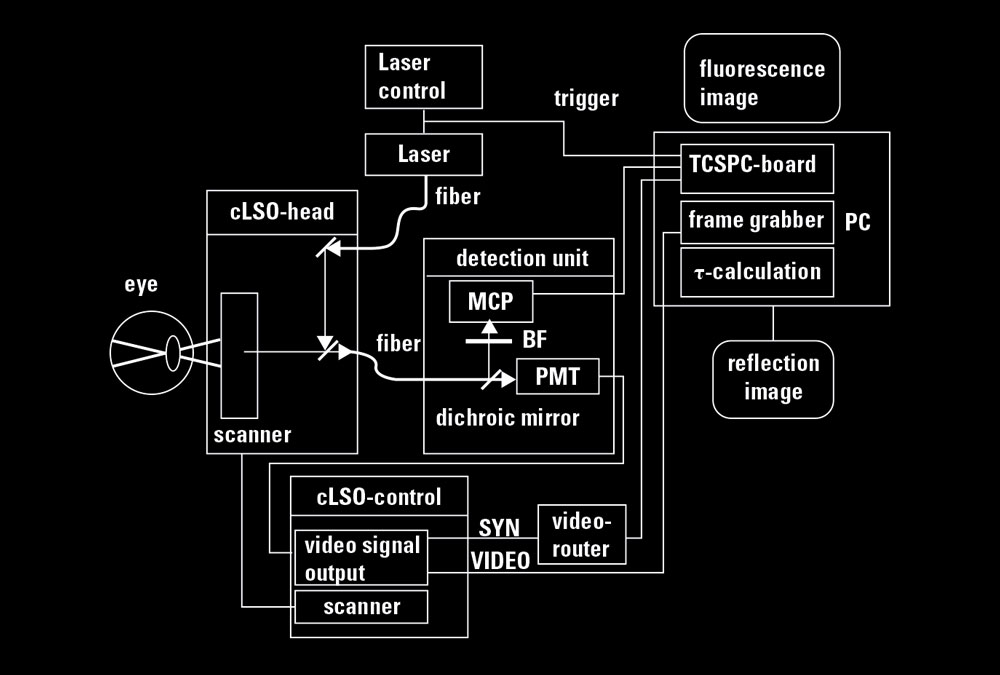
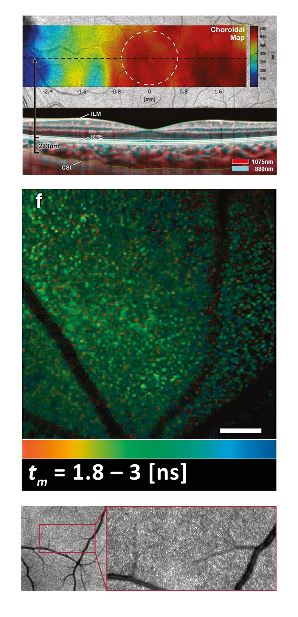
First prototype device for Fluorescence Lifetime Imaging Ophthalmoscopy (FLIO)
Wolf, Dysli, et al.
First commercial version of objective measurement of the BMO-MRW with APS: GMPE
Swept-source OCT prototype for anterior segment imaging
HEYEX 2 – Next generation image management platform
⇩
2015
First in vivo two-photon FA on animals
Patent granted for use of phase plates for abberation reduced imaging
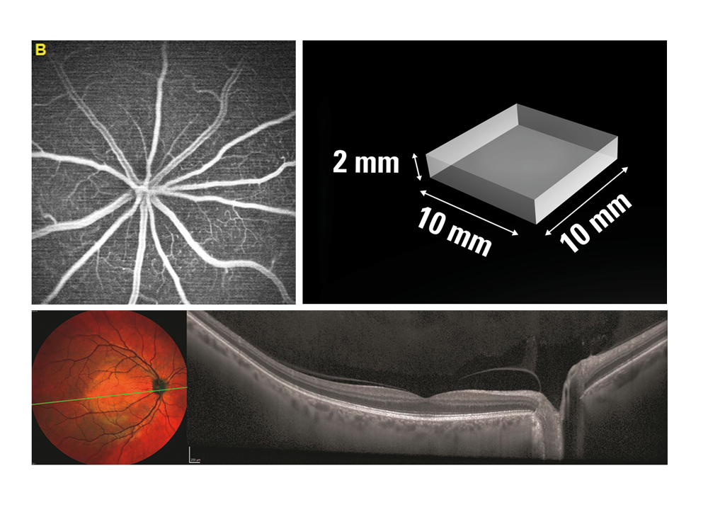
First simultaneous widefield OCT and widefield cSLO imaging
⇩
2016
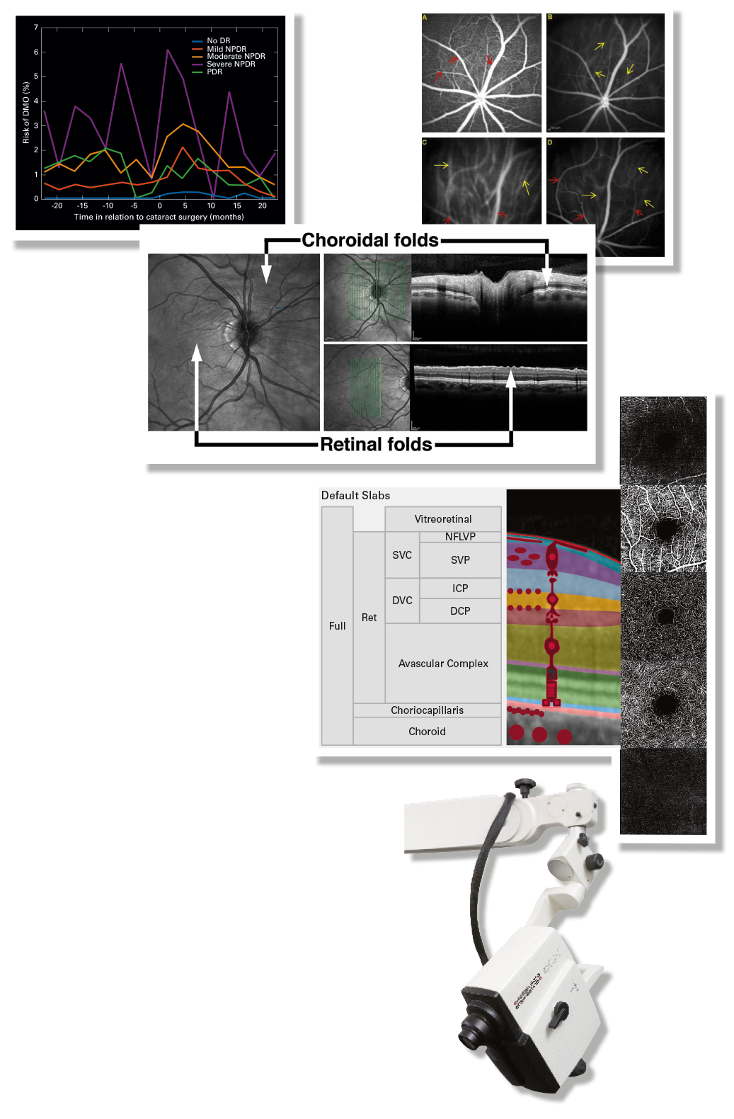
First OCT Angiography with full axial resolution, real-time eye-tracking, AutoRescan and dynamic visualization with Hybrid Angiography
Real-time swept-source OCT imaging in cataract and refractive surgery (VICTUS, B+L)
⇩
2017
Real-world data mining through structured EMR
Denniston, et al.
First in vivo two-photon imaging of retina (FA and ICGA) in rodents
Jayabalan, et al.
NASA Space Associated Neuro Ocular Syndrome study (SANS) – OCT2 in space
Lee, Mader, et al.
Scrolling through the enface OCTA images
First mobile OCT with real-time eye tracking (FLEX) – Aqueous angiography study
Huang, et al.
⇩
 |
2018Modified SPECTRALIS HYDRA in Switzerland / Moorfields Lab system combining twophoton imaging with FLIO High Magnification Module |
⇩
2019
First combination of High Magnification Imaging with customized phase plates
Holz, et al.
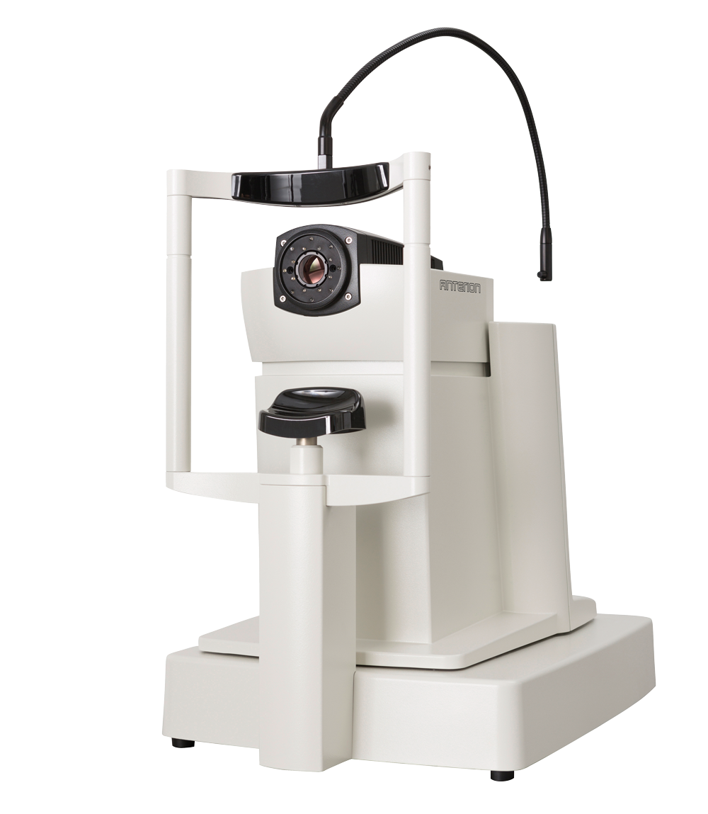
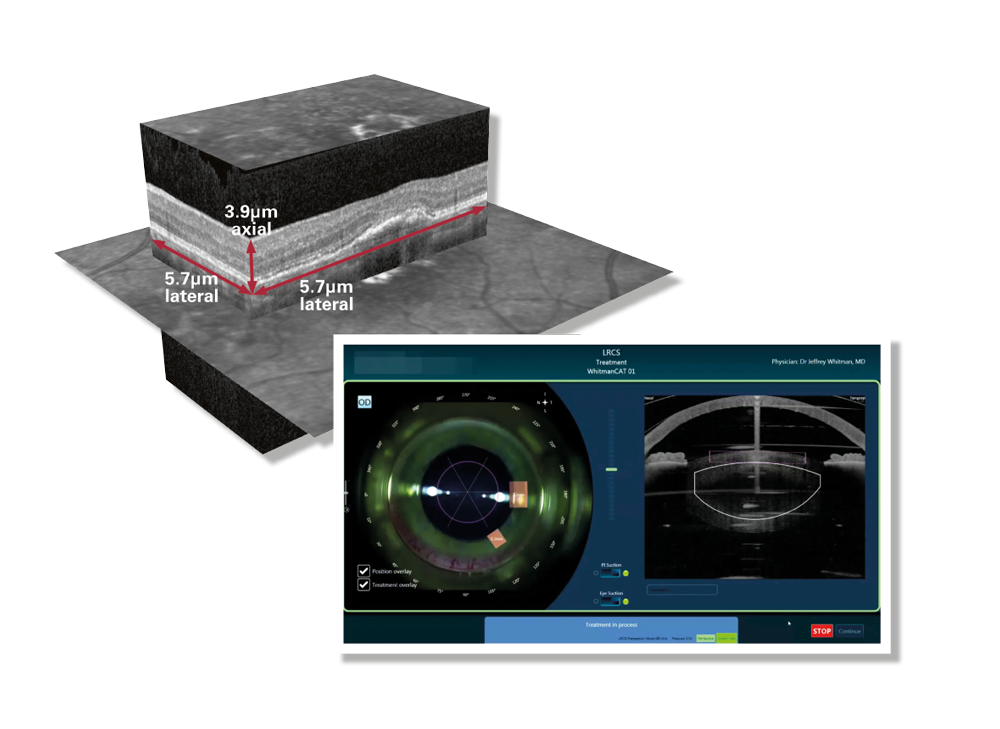
Swept-source OCT multimodal biometric system for anterior segment (ANTERION)
⇩
2020
First commercial prototype for high resolution OCT
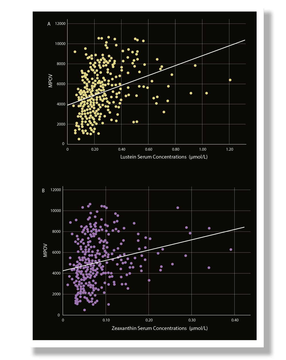
Macular Pigment Optical Volume (MPOV), standardized measurement of macular pigment
Nolan, et al.


