SPECTRALIS – Proven Imaging Platform for Diagnostic Hubs
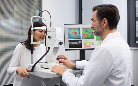
The SPECTRALIS imaging platform is being utilised at diagnostic hubs across the country to offer more people with sight threatening conditions timely treatment when pressures on services due to the pandemic have caused delays. The SPECTRALIS is ideally suited to the diagnostic hub environment with its exceptional image quality, small footprint, multimodal imaging capabilities, up to one micron reproducibility, and ease of use.
Ease of use
The SPECTRALIS OCT model configuration is a “slimline” platform that features a smaller headrest and a joystick trigger button, for fast image capture and a reduced footprint compared to the traditional SPECTRALIS HRA or HRA+OCT models. The joystick control is designed for ease of use, and makes capturing high-quality images simple, even for inexperienced users. The "slimline" SPECTRALIS is an affordable solution that can be integrated into the existing hospital infrastructure.
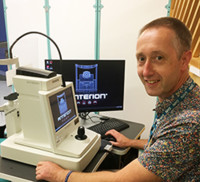
“The OCT is easy to use and allows a high degree of flexibility in imaging. The manual focus and joystick controls are very useful in an NHS clinic as we are seeing patients that can be traditionally challenging to image. The OCT really makes the difference in this regard and allows us to capture images, even in patients with nystagmus or high myopia”.– Richard Bell, Senior Medical Photographer, The Royal Victoria Infirmary.
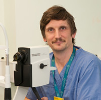
“We are finding some patients had previously been scanned unsuccessfully on other machines, but we have now obtained images using the SPECTRALIS. The doctors couldn’t believe how good the images were, given that they had been acquired by staff who had only been in the job for two months.”– Peter Holm, Co-ordinator & Practice Educator for Ophthalmic Imaging, Moorfields Eye Hospital
High-resolution multimodal imaging
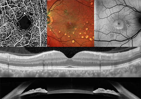
All SPECTRALIS are modular and upgradeable, allowing you to configure the platform to your individual requirements and budget, whilst future-proofing the technology in clinic for many years to come. The combination of modalities on one platform aids the clinic in working through the backlog of overdue appointments efficiently, whilst maintaining the clinical sensitivity and specificity required for delivering diagnostic confidence and improved patient care.
“When choosing an OCT, it was all about opting for high-quality, high-resolution images. As the patient isn’t going to be seeing the doctor face-to-face, the appointment at the hub is 100 percent diagnostic focused. The OCT images are imperative for the doctor to be able to make a diagnosis and assign the right treatment pathway, so image quality and detail is everything. For me personally, the resolution of the SPECTRALIS and its excellence in imaging highly myopic patients made it the first choice”
– Peter Holm, Co-ordinator & Practice Educator for Ophthalmic Imaging, Moorfields Eye Hospital
Technologies designed for the reality of imaging patients
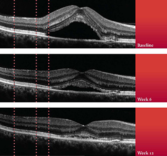
TruTrack Active Eye Tracking, inherent in the DNA of every SPECTRALIS, mitigates for eye movement during scanning, enabling the operator to acquire high-quality images independent of blinks and motion artefacts. Additional clinical benefits of TruTrack Active Eye Tracking are precise, automated retinal follow-up scanning, retinal thickness measurement reproducibility to 1 micron, and excellent image quality throughout the volume scan. This superior performance allows a more effective management of patients, even in challenging cases.
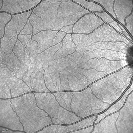
SPECTRALIS confocal scanning laser ophthalmoscopy (cSLO) imaging minimises the effects of light scatter and can be used to acquire images even in patients with cataracts. As there is no bright flash when using infrared imaging, scans can be acquired with no pupil dilation in any lighting conditions, which is highly desirable in a fast-track diagnostic hub environment.
“We don’t dilate the eyes of patients who attend the glaucoma hub, which is no problem for the OCT. This speeds up the workflow and encourages more patients to attend their appointment as they are able to drive”.
– Richard Bell, Senior Medical Photographer, The Royal Victoria Infirmary.
“For me, the SPECTRALIS is by far the best OCT for imaging patients with small pupils. We have the ability to see around cataracts and get a really good image with little dilation.”
– Peter Holm, Co-ordinator & Practice Educator for Ophthalmic Imaging, Moorfields Eye Hospital
Read the full interviews with Richard Bell, Senior Medical Photographer, The Royal Victoria Infirmary, and Peter Holm, Co-ordinator & Practice Educator for Ophthalmic Imaging, Moorfields Eye Hospital, about how SPECTRALIS is facilitating access to timely treatment for patients at diagnostic hubs at their NHS trust.
Contact Heidelberg Engineering for further information about configuring a SPECTRALIS for your diagnostic hub or clinic.
For more valuable information about our products, educational offerings, learning materials and events
Subscribe to our Newsletter orContact us
