Improve your diagnostic capabilities with Glaucoma Module Premium Edition and Nsite Analytics
Early diagnosis and monitoring
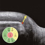
GMPE provides a comprehensive analysis of the optic nerve head, retinal nerve fiber layer, and ganglion cell layer by precisely matching unique scan patterns to the fine anatomic structures relevant in glaucoma diagnostics. GMPE can be combined with Nsite Analytics Module, which allows the clinician to make a distinction between glaucoma and other neurodegenerative diseases, such as multiple sclerosis, and manage the patient accordingly. GMPE and Nsite flag even very small deviations to allow for early diagnosis and monitoring.
Precise and objective measurement visit after visit
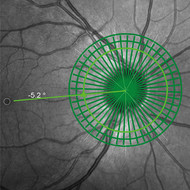
Crucially, GMPE and Nsite use a proprietary Anatomic Positioning System (APS) to remove operator variability during scanning by creating an anatomic map of each patient’s eye so that all scan protocols are automatically oriented according to the patient’s individual anatomy. This allows a highly sensitive assessment of structural change when combined with the AutoRescan function, which automatically places follow-up scans in precisely the same position visit after visit. Studies have shown that SPECTRALIS with AutoRescan technology can reliably measure changes in retinal nerve fiber layer thickness as small as 1 micron, providing the clinician with confidence in the data obtained by their device.
Fast image acquisition with OCT2
Combine the GMPE and Nsite modules with OCT2 Module and enjoy a high scan rate of 85,000 Hz for fast image acquisition speeds to improve clinic workflow. The OCT2 Module can be added to your existing upgradeable SPECTRALIS instrument so you can retain current patient data for continued follow-up to measure change over time.
Heidelberg Engineering at UKEGS in Cheltenham
Visit Heidelberg Engineering at stand 9 to discuss how SPECTRALIS can improve patient care in your clinic with one of our expert imaging consultants on the 17th and 18th November 2016 at the UK and Eire Glaucoma Society (UKEGS) Meeting in Cheltenham.
| The Window2Brain website introduces how Nsite Analytics adds new dimensions to understanding axonal loss by imaging and measuring the RNFL in a fast, non-invasive technique. |
| The Glaucoma Module Premium Edition PDF Tutorial supplies comprehensive explanations about the physiological and technical background, the correct Bruch‘s Membrane Opening (BMO) positioning, the available scan patterns and the interpretation of the obtained data. |


