 Heidelberg, Germany – Heidelberg Engineering, the industry leader in ophthalmic diagnostic imaging, announces the reintroduction of its HRT3 Rostock Cornea Module (RCM) to offer in vivo corneal confocal microscopy in its anterior segment product portfolio, supporting cornea specialists worldwide to further improve patient care.
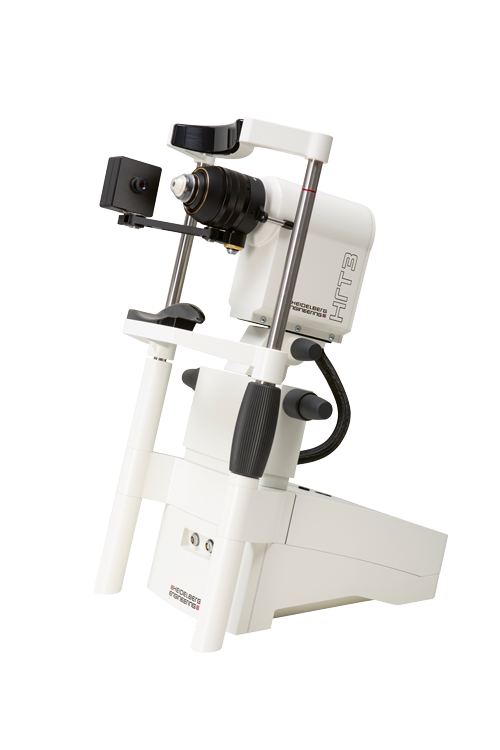
HRT3 RCM is a compact ophthalmic device that uses confocal scanning laser microscopy to provide high-resolution images of the cornea, the conjunctiva or the limbus at the cellular level. It capitalizes on the proven technology that has differentiated Heidelberg Engineering as an innovator in both the posterior and the anterior segment market for 30 years. The company collaborated with the research group of Prof. Dr. Rudolf Guthoff at the University of Rostock, Germany, to develop a dedicated lens and software application for the HRT platform that enables in vivo microscopy of the cornea.
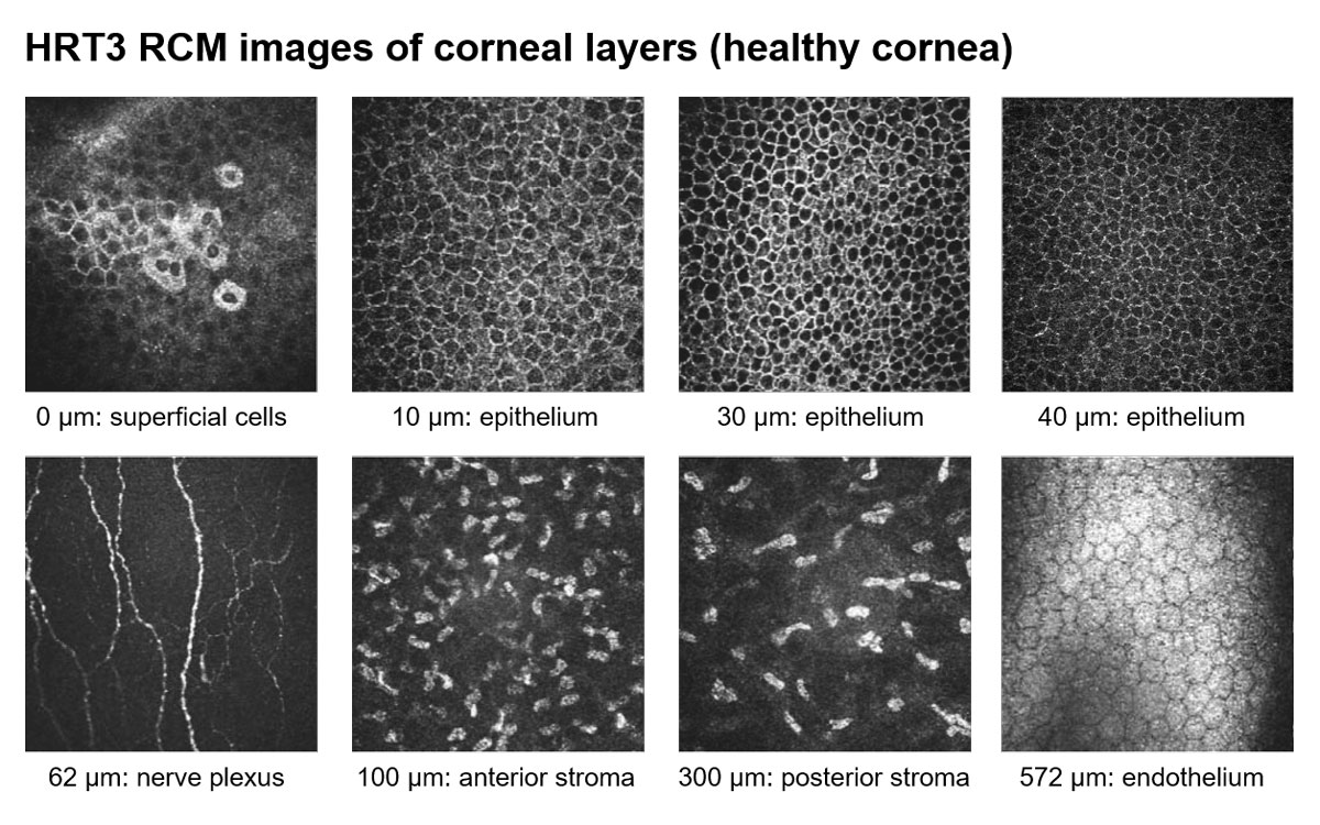
The resulting HRT3 RCM provides unique en face images of corneal cells and structures. It scans the cornea with a field of view of up to 400 x 400 μm, resulting in high-resolution images that allow the user to navigate through all corneal layers – including the subdifferentiation of various epithelial cell layers –, identify keratocytes subpopulations and even visualize the corneal subbasal nerve plexus. This comprehensive in vivo assessment of the cornea can aid clinicians in the diagnosis and monitoring of corneal pathologies, pre- and post-surgery evaluation, the assessment of dry eye disease, and in the evaluation of corneal nerve structure in diabetic patients.
Its unprecedented imaging capability has made the HRT3 RCM an ideal device for scientific research, which has resulted in numerous publications1. HRT3 RCM has also proven its relevance as a diagnostic platform for daily clinical practice. With growing demand from the clinical evidence base and global market, Heidelberg Engineering decided to reintroduce the in vivo microscope as part of its commercial portfolio.
“The relaunch of the HRT3 RCM is exciting as this unique imaging device aids the ophthalmologist in the diagnosis of corneal diseases by allowing imaging at a cellular level. The high-resolution images have been pivotal for visualization of corneal infections such as acanthamoeba and fungi, as well as quantification of corneal nerves in patients with diabetes mellitus and dry eye disease. The HRT3 RCM is a very promising tool for both the clinician wanting to make a diagnosis in the clinic, as well as researchers interested in the cellular basis of disease, thus facilitating in vivo histology”, says Dr. Jaya Chidambaram, BSc (Hons), MBBS, MRCOphth, PhD, Senior Lecturer, University of Manchester, United Kingdom.
HRT3 RCM will be relaunched in spring 2020, including a new headrest for corneal assessment. It will also integrate into the company’s HEYEX 2 image management software environment, which enables new functionalities for streamlined workflows, such as automated report generation and archiving, or drag-and-drop exports for paperless data sharing.
“The new ‘European Medical Device Regulation’ will compel device manufacturers to re-evaluate their product portfolios: For some legacy products, the cost of adapting to the new regulation may not be justifiable. In addition to the contribution the HRT3 RCM has made to scientific research, there is an unmet clinical need for high-resolution in vivo corneal microscopy. We have decided to renew the HRT3 RCM both to advance science and improve patient care,” explains Erich E. Bangert, Vice President of Sales & Service, Heidelberg Engineering.
With its ability to investigate the cornea at the cellular level, HRT3 RCM is expected to empower clinicians in fast-growing clinical areas such as dry eye disease, infectious corneal diseases, corneal dystrophies, diabetic neuropathy, as well as refractive and corneal treatment surgery.
1 Villani E, Baudouin C, Efron N, Hamrah P, Kojima T, Patel S V, Pflugfelder S C, Zhivov A, Dogru M. “In Vivo Confocal Microscopy of the Ocular Surface: From Bench to Bedside”. Current Eye Research. 2014; 39(3). Link: https://www.ncbi.nlm.nih.gov/pmc/articles/PMC3960287/


