Pioneering research from the University of Edinburgh is providing vital insights into kidney health using Optical Coherence Tomography (OCT) scans of the retina and choroid. Professors Neeraj (Bean) Dhaun, Professor of Nephrology; Honorary Consultant Nephrologist; Director Edinburgh Clinical Research Facility and Matt Bailey, Professor of Renal Physiology, lead the team at Edinburgh and have published multiple papers that show non-invasive OCT imaging provides a “window to the kidney”.
“Currently, kidney health is assessed using blood tests or invasive kidney biopsies, which are not patient friendly”, says Professor Dhaun. “Also, performing multiple biopsies over time to monitor kidney function can be uncomfortable for patients and carries some risk. There is a desire for gentler methods to track disease progression or treatment response, and to predict future health issues.”
The eye and the kidney share structural similarities, and their disease pathways are also alike. This means some diseases might manifest similarly in both organs, allowing researchers to glean insights into kidney function by examining the eye. The team at Edinburgh used OCT to show an association between kidney function and the thickness of the retina and choroid.
“We compared OCT scans from healthy volunteers and those from patients at varying stages of chronic kidney disease (CKD) and found a relationship”, says Professor Dhaun. “Patients with CKD had thinner retinal and choroidal layers compared to healthy individuals, and these layers became progressively thinner as kidney function deteriorated”.
Remarkably, as early as one week after a kidney transplant, patients previously suffering from kidney failure showed thicker retinal and choroidal layers, which continued to thicken for at least a year as their kidney function improved. On the other hand, healthy individuals who donated a kidney experienced gradual thinning of these layers over the year following their donation.
Imaging patients with CKD initially presented some challenges to the research team.
“Patients who take part in the research need to have OCT imaging at certain time intervals of their disease stage and treatment. They attend a variety of clinics across the hospital site including outpatients, the dialysis unit, high dependency and intensive care units. Some patients who require imaging are in recovery, for example after a kidney transplant, and need to lie flat during this time,” says Professor Dhaun.
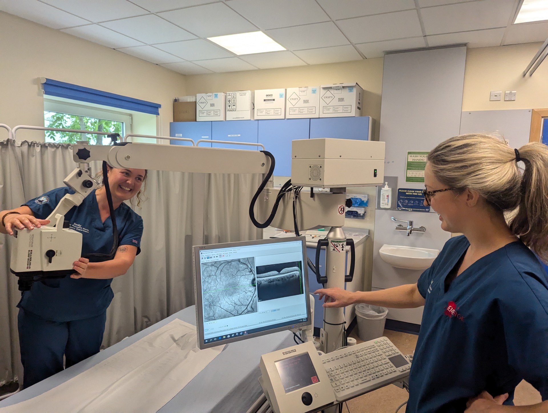
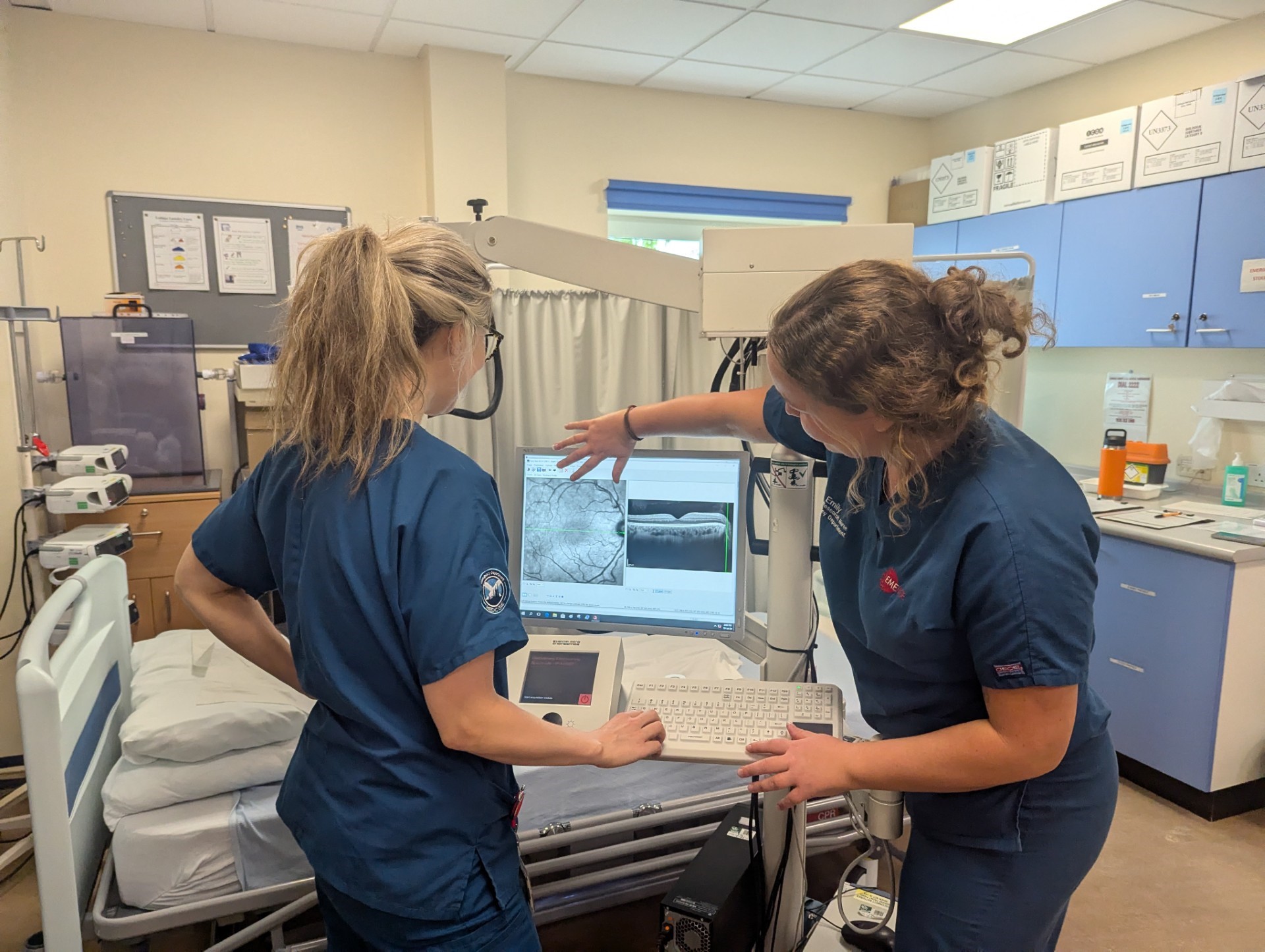
Traditional OCT devices are not designed to move around the hospital freely or have the capability to image patients in the supine position. The challenge was overcome by using the Heidelberg Engineering SPECTRALIS with Flex Module.
“The SPECTRALIS Flex is on wheels and transportable, so we can move the device around the hospital to the various clinics or ward areas. It is easier and more efficient to bring the device to wherever the patient was being seen or treated in hospital than to try to bring the patient to the device, such as when a patient is having dialysis for example,” explains Professor Dhaun. “Because the SPECTRALIS Flex has a moveable base with an extending flexible arm it also meant we could take OCT images of eyes in patients in recovery who need to lie flat.”
The SPECTRALIS with Flex Module utilises the same core technologies and multimodality imaging capabilities of the regular table-mounted SPECTRALIS, which is used globally in ophthalmology clinics.


“The TruTrack eye tracking and AutoRescan facilities enable you to do a baseline scan and then subsequent scans at the exact same location in the retina; this was critical to this research”, says Professor Dhaun. “With one device we can do retinal nerve fiber layer (RNFL), macular volume scans, and OCT with Enhanced Depth Imaging (EDI) enabled for detailed visualization of the choroid. We also did OCT Angiography (OCTA) imaging and are yet to analyse those images to see what they tell us.”
The researchers have also demonstrated that in CKD patients, the thickness of the retina and choroid could predict future decline in kidney function. The team is now investigating the precise mechanisms causing these ocular changes in relation to kidney function changes.
“OCT measurements of the retina and choroidal layers could serve as a non-invasive, sensitive method for monitoring kidney health. This technique might also help predict which patients are at risk of further health issues, such as cardiovascular disease, enabling earlier intervention and treatment”, explains Professor Dhaun. “We are now exploring the utility of these OCT measures as potential clinical biomarkers and as short- and medium-term endpoints in clinical trials. We hope measuring changes in the retina might indicate whether – and in what way – the kidney responds to potential new treatments and that this research will aid in the development of new therapies and better patient outcomes”
Find out more about the research at Edinburgh here. Information about the SPECTRALIS with Flex Module can be found here.


