Discover retinal details at the microstructural level with SPECTRALIS
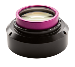
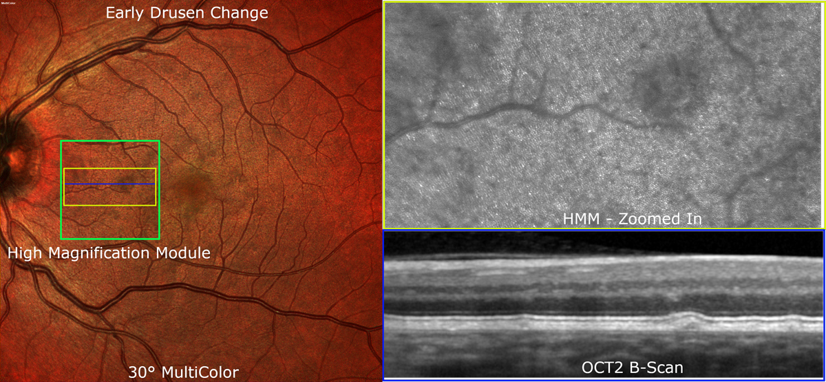
 The SPECTRALIS® High Magnification Module (HMM) elegantly demonstrates the capability of confocal scanning laser ophthalmoscopy (cSLO) to resolve ocular microstructures by reducing intraocular light scattering, even in eyes with cataracts. Far from being a simple digital zoom, the HMM visualizes significantly more microstructural detail of the ocular fundus than is possible with a standard SPECTRALIS lens.
The detail you see in HMM images may provide novel insights into the pathogenesis and progression of retinal diseases, providing you with further possibilities to refine surgical and treatment regimens.

Visit us at the Annual Meeting of ARVO (April 28th - May 2nd), at booth #1117, to experience the SPECTRALIS High Magnification Module for yourself.


