Christmas edition of highly regarded imaging symposium held in Vienna
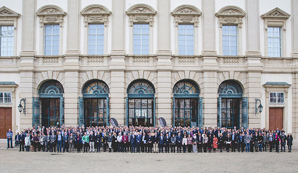
Hemel Hempstead, UK – 349 participants from 44 countries attended the 14th International SPECTRALIS Symposium (ISS) on the 25-26th November at the Garden Palace Liechtenstein in Vienna, Austria.
“Participants enjoyed lectures devoted to cutting-edge research and clinical applications using diagnostic imaging tools for retina and glaucoma from some of the world’s most eminent ophthalmic professionals”, said Christopher Mody, Director of Clinical Services. “Highlights of the programme included updates on autofluorescence, OCT angiography normal anatomy and pathology and the latest innovations in glaucoma detection and progression.”
The ISS included an evening at the renowned Spanish Riding School in Hofburg palace, a magnificent baroque building built in 1735, including a visit to the stables to enjoy a traditional outdoor Christmas Market, set up exclusively for ISS guests.
Save the date for the 15th International SPECTRALIS Symposium, taking place in Boston, USA, on the 20th and 21st October 2017. Visit www.HE-Academy.com to find over 100 hours of education on offer from Heidelberg Engineering in 2017. 349 participants from 44 countries attended the 14th International SPECTRALIS Symposium
OCT Angiography Module for SPECTRALIS now available
The SPECTRALIS OCT Angiography Module non-invasively produces detailed, three-dimensional representations of the perfused retinal and choroidal vasculatures.Heidelberg Engineering has started delivering the OCT-Angiography Module to SPECTRALIS® customers outside the United States. The OCT Angiography Module non-invasively produces detailed three-dimensional representations of the perfused retinal and choroidal vasculatures.
Heidelberg, Germany – The SPECTRALIS® expandable diagnostic imaging platform can be upgraded with the OCT Angiography Module to perform non-invasive, layer-by-layer examinations of flow in the vascular networks of the retina and choroid.
The OCT Angiography Module can be added to new and existing upgradeable SPECTRALIS devices with the OCT2 Module. The multimodal imaging platform allows clinicians to compare OCT angiographies to other modalities such as structural OCT and dye-based angiographies as well as infrared, MultiColor and BluePeak images, dependent on the SPECTRALIS model.
“In combination with other imaging modalities, OCT angiography enables a more comprehensive understanding of vascular abnormalities. The SPECTRALIS truly integrates OCTA with other clinically well-established imaging modalities to facilitate for instance direct, pixel-to-pixel OCTA follow-ups on an existing FA,” said Dr. Kester Nahen, Managing Director of Heidelberg Engineering.
High resolution for precise vascular details
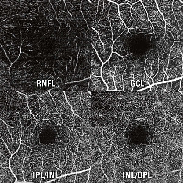
SPECTRALIS offers high resolution OCT angiography with a lateral 5.7 microns per pixel for visualization of capillaries. An axial resolution of 3.9 microns per pixel enables precise multilayer segmentation. All four vascular plexuses of the retina as described by Tan et al.1 (located in the nerve fiber layer, in the ganglion cell layer, at the border of the inner plexiform layer and inner nuclear layer, and at the border of the inner nuclear layer and outer plexiform layer) can be investigated. Like all SPECTRALIS diagnostic imaging modalities, the OCT Angiography Module benefits from the precision of TruTrack Active Eye Tracking which avoids motion artifacts and ensures high resolution images.
1 Priscilla Ern Zhi Tan, Invest Ophthalmol Vis Sci. 2012;53:5728–5736
Dr. Gerhard Zinser receives Founders’ Award of the Optometric Glaucoma Society
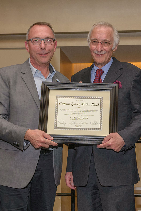
Dr. Gerhard Zinser (right) receives the Founders’ Award from Dr. John Flanagan, one of the founders of the Optometric Glaucoma Society. Photo: Andie Petkus Photography.
Heidelberg, Germany – Dr. Gerhard Zinser, co-founder and Managing Director of Heidelberg Engineering, is the first non-clinician to be honored with this prestigious award. Earlier this year he was also elected as part of The Ophthalmologist’s Power List of the top 100 most influential people in ophthalmology.
The Founders’ Award was handed over by Dr. John Flanagan, one of the founders of the Optometric Glaucoma Society, during the society’s Annual Scientific Meeting in Anaheim, CA, on November 8th.
In his laudatory speech, John Flanagan, OD, PhD, (Dean School of Optometry, UC Berkeley, CA) emphasized Dr. Zinser’s significant and long-standing contribution to diagnostic imaging in eye-care with innovative and reliable devices which have turned into household names for eye-care specialists worldwide. Dr. Flanagan summarized the achievements of Dr. Zinser and Heidelberg Engineering dating back to the launch of the first diagnostic imaging device geared towards glaucoma diagnostics in 1991, the Heidelberg Retina Tomograph (HRT). Dr. Flanagan reminded the audience also of the tremendous impact on eye-care of the most current contribution of Heidelberg Engineering, the multimodal SPECTRALIS imaging platform.
“I am honored to receive this award and accept it in the name of the whole Heidelberg Engineering team”, said Dr. Gerhard Zinser.
Since the establishing of the Optometric Glaucoma Society, the Founders’ Award has only been awarded to three clinician scientists, all of them glaucoma research luminaries: Douglas R. Anderson, MD (Bascom Palmer, Miami, FL) received the honor in 2011, Robert N. Weinreb, MD (Shiley Eye Institute, UC San Diego, CA) received the honor in 2012 and Harry A. Quigley, MD (Johns Hopkins, Baltimore, MD) in 2015.
Power List 2016
Earlier this year the readers of the journal The Ophthalmologist voted for Dr. Gerhard Zinser to be part of its Power List 2016, as one of the top 100 most influential people in the world of ophthalmology for the second consecutive time. According to a nominator quoted by The Ophthalmologist: “He is responsible for several of the major diagnostic imaging innovations in ophthalmology. Further, he is actively involved in cutting-edge research and in sponsoring innovative activities that will translate into major products in the coming decade.”
For the employees and distribution partners of Heidelberg Engineering, the recognition awarded to Dr. Zinser’s achievements is an encouragement to continue bringing innovative diagnostic devices and IT solutions to eye-care specialists around the world.
About the Optometric Glaucoma Society (OGS)
The Optometric Glaucoma Society´s mission is to encourage excellence in care of glaucoma patients through education and scientific investigation. The main goals of the OGS are to promote the education of optometrists, the acquisition of new knowledge about glaucoma and much more.
Next generation HEYEX image management platform piloted in the UK
Hemel Hempstead, UK – Next generation image management software for the SPECTRALIS imaging platform is to be piloted at several key sites across the UK.
The new Heidelberg Eye Explorer (HEYEX) platform is a comprehensive ophthalmic image management and device integration solution for the clinic. Its flexible and modular design provides a solution to clinics of all sizes, from small practices with a single instrument to large enterprises with client server architecture, numerous instruments and multiple sites.
“The HEYEX platform will provide the reliability, security and quality required to improve patient care in the most demanding clinical environments. It offers rapid image acquisition and viewing for improved clinic workflow”, explains Kenny Boyle, Key Accounts Manager at Heidelberg Engineering
The first installation as part of the pilot programme occurred at Eyecare Medical in Macclesfield in September with installations at other key sites nationwide scheduled for 2017.
Heidelberg Engineering to bring OCT to life nationwide
Hemel Hempstead, UK – Heidelberg Engineering is once again hitting the road to bring OCT to life with OCT LIVE: a series of CET-accredited educational events nationwide.
Delegates will be guided through the patient journey, from scanning the patient live in HD on the big screen to interpreting the images and making a decision on patient management. These interactive, CET-accredited sessions challenge the classic case study book format and demonstrate how a multimodal imaging approach can be used in real life to pinpoint pathology and refer with confidence.
“The OCT LIVE roadshows are an exciting way for practitioners to learn about OCT as they can see the scan taking place live in high definition and learn how they can use OCT for the detection and monitoring of retinal disease and optic neuropathies” explains Christopher Mody, Director of Clinical Services at Heidelberg Engineering.
The roadshows are held in the evening, starting at 6.30pm and are suitable for OCT beginners and skilled users alike. Three interactive CET points are available for both optometrists and dispensing opticians.
OCT Angiography Module for SPECTRALIS benefits from superior resolution
SPECTRALIS Hybrid AngiographyHemel Hempstead, UK – The OCT Angiography (OCTA) Module for the SPECTRALIS diagnostic imaging platform is now available to order and will begin shipping in November 2016. The OCTA Module uses a complex mathematical model fed by full spectrum OCT data. The full spectrum approach enables three-dimensional OCTA imaging with high axial resolution separating thin vascular plexuses.
“With a lateral resolution of 5.7 microns and an axial resolution of 3.9 microns we are able to resolve the individual vascular capillary plexus within the retina and provide very high resolution images for the clinician. This technology can then be combined with conventional fluorescein and ICG angiography to form hybrid angiography, which provides pixel by pixel correlation between OCTA and dye-based angiographies as well as infrared, MultiColor and BluePeak images”, explained Christopher Mody, Director of Clinical Services.
The OCT Angiography Module can be added to new and existing upgradeable SPECTRALIS devices with the OCT2 Module. Like all SPECTRALIS diagnostic imaging modalities, the OCT Angiography Module benefits from the precision of TruTrack Active Eye Tracking, which avoids motion artifacts and ensures high resolution images.
Heidelberg Engineering sets the standard in OCT training for dispensing opticians
Hemel Hempstead, UK – Heidelberg Engineering offers a diverse range of OCT training courses, all of which are accredited for both optometrists and dispensing opticians. The company has already run more than 50 points worth of interactive CET in 2016, with plans to increase the number of courses in 2017. Courses focus on fundus and OCT image acquisition and analysis for both retinal diseases and glaucoma. Many use the OCT LIVE format, where patients are imaged live on the big screen followed by a discussion on the use of imaging in diagnosis and referral protocol.
“We would like to highlight that our training courses are accredited for dispensing opticians too. Despite acquiring images with OCT routinely, dispensing opticians are often overlooked when it comes to CET points for imaging.”, explains Christopher Mody, Director of Clinical Services.
Visit www.HE-Academy.com to find out more about upcoming events and to book your place.
SPECTRALIS OCT Angiography Module to be presented at AAO
Heidelberg Engineering is showcasing the OCT Angiography (OCTA) Module for its SPECTRALIS® diagnostic imaging platform at the American Academy of Ophthalmology Annual Meeting in Chicago. The OCT Angiography Module non-invasively produces detailed three-dimensional representations of the perfused retinal and choroidal vasculatures.
Heidelberg, Germany – The SPECTRALIS® expandable diagnostic imaging platform can be upgraded with the OCT Angiography Module to perform non-invasive, layer-by-layer examinations of flow in the vascular networks of the retina and choroid. Combined with structural OCT and fluorescence angiography, the OCT Angiography Module enables a more comprehensive understanding of vascular abnormalities.
The OCT Angiography Module can be added to new and existing upgradeable SPECTRALIS devices with the OCT2 Module. The multimodal imaging platform allows clinicians to compare OCT angiographies to other modalities such as structural OCT and dye-based angiographies as well as infrared, MultiColor and BluePeak images, dependent on the SPECTRALIS model.
High resolution for precise vascular details
SPECTRALIS offers high resolution OCT angiography with a lateral 5.7 microns per pixel for visualization of capillaries. An axial resolution of 3.9 microns per pixel enables precise multilayer segmentation. All four vascular plexuses of the retina as described by Tan et al.1 (located in the nerve fiber layer, in the ganglion cell layer, at the border of the inner plexiform layer and inner nuclear layer, and at the border of the inner nuclear layer and outer plexiform layer) can be investigated. Like all SPECTRALIS diagnostic imaging modalities, the OCT Angiography Module benefits from the precision of TruTrack Active Eye Tracking which avoids motion artifacts and ensures high resolution images.
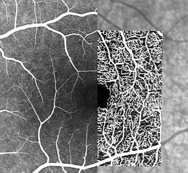
Hybrid angiography
Hybrid angiography with SPECTRALIS high-resolution OCT Angiography Module identifies vascular detail and correlates it with dye-based angiography. Left to right: FA, OCTA and IR images.
The SPECTRALIS platform offers a unique combination of non-invasive OCT angiography with dye-based, gold-standard fluorescein (FA) or indocyanine green (ICGA) scanning laser angiography. With this hybrid angiography approach, OCTA images can be directly correlated pixel to pixel to FA or ICGA, setting a baseline for subsequent OCTA-only follow-up examinations. The Scan Planning Tool automates OCTA scan placement on a region of interest identified on FA/ICGA or any other previously-acquired cSLO image. Such reference images can originate from the same or another SPECTRALIS device. With the convergence of established and emerging imaging techniques, the SPECTRALIS platform supports the understanding and adoption of new diagnostic tools such as OCTA in routine clinical practice.
“There is a great deal of interest in OCTA and at the same time uncertainty due to its technical limitations and interpretation challenges. We see multimodal imaging as a good approach to deal with those issues. For instance, OCTA is a valuable imaging modality to follow up on an existing FA, and our SPECTRALIS platform offers the possibility to do exactly that,” said Dr. Kester Nahen, Managing Director of Heidelberg Engineering.
OCTA technology and SPECTRALIS
The OCTA module of the SPECTRALIS uses a complex mathematical model fed by full spectrum OCT data. The full spectrum approach enables three-dimensional OCTA imaging with high axial resolution separating thin vascular plexuses. The mathematical model is highly sensitive and allows the visualization of small changes between consecutive OCT scans representative of vascular flow. This method results in high contrast between areas of vascular flow and surrounding tissue.
The SPECTRALIS is an ophthalmic imaging platform with an upgradable, modular design. This platform allows clinicians to configure each SPECTRALIS to the specific diagnostic workflow in the practice or clinic. Options include: OCT, multiple scanning laser fundus imaging modalities, widefield and ultra-widefield modules, scanning laser angiography and OCT angiography.
Availability
The OCT Angiography Module is not for sale in the United States. Heidelberg Engineering will deliver the OCTA Module to customers outside the U.S. in November 2016.
1 Priscilla Ern Zhi Tan, Invest Ophthalmol Vis Sci. 2012;53:5728–5736
Leading international glaucoma specialist confirmed to present at 100% Optical 2017
Sanjay Asrani, Professor of Ophthalmology and Clinical Director of the Duke Eye CentreHemel Hempstead, UK – Highlighting the world-class connections that Heidelberg Engineering engenders, their guest speaker at 100% Optical 2017 has been confirmed as Sanjay Asrani, Professor of Ophthalmology and Clinical Director of the Duke Eye Centre, North Carolina, USA.
The CET-accredited lecture he will deliver at the show will reflect his position as a leading international glaucoma specialist and American Academy of Ophthalmology Award winner.
“Heidelberg Engineering are delighted to be welcoming Professor Asrani back to the UK to speak at 100% Optical once again”, explains Christopher Mody, Director of Clinical Services. “His show-stopping presentation on the main stage in 2015 ‘MultiColor + OCT: The Interface of Retina and Glaucoma‘ was extremely well received and his next instalment at the 2017 meeting will showcase the latest multimodal imaging techniques for the diagnosis and management of glaucoma in practice”.
Professor Asrani’s lecture will take place at 12.00pm on Sunday 5th February and registration for the lecture will open on the 100% Optical website soon.
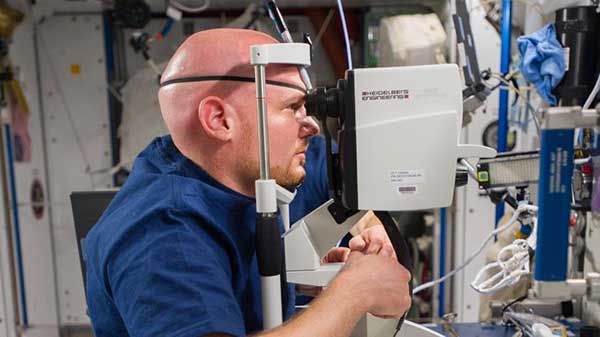
Eye scanning technology used by NASA to monitor vision in astronauts aboard the International Space Station is now available in High Street Opticians
Hemel Hempstead, UK – When astronauts spend lengthy periods of time in space, they can experience changes in their vision believed to be caused by space travel and micro-gravity. These changes are not only a concern for today’s astronauts on board the International Space Station (ISS), but for astronauts that will participate in human missions to Mars lasting up to 3 years in the future.
Evidence suggests that during prolonged periods in space, the extreme environment could do irreversible damage to an astronaut’s eyes. NASA has commissioned a comprehensive study in the hope that in-flight monitoring and timely treatment will be able to counter the detrimental visual effects of living in space.
Astronauts undergo a variety of eye examinations at regular time intervals during their mission. One of the technologies that is being used aboard the International Space Station measures very small changes in the structure of the eye. The SPECTRALIS is a high-tech eye scanner that employs eye tracking technology to scan in precisely the same location every time, detecting as little as 1 micron of change. NASA uses the SPECTRALIS to perform a precise, objective examination at baseline prior to space flight, at routine intervals during missions and following space travel. It is thought that the SPECTRALIS could be an on-board “early-warning system”, detecting small changes before astronauts are aware of deterioration in their vision.
Having once been exclusive to specialists and researchers, this amazing eye scanning technology is now available to High Street opticians. With just over 100 centres nationwide, the SPECTRALIS eye health check, performed in seconds and without any pain or sensation, can highlight the earliest signs of disease – long before any symptoms appear.