EssilorLuxottica signs agreement to acquire a majority stake in Heidelberg Engineering and continues to make moves in the med-tech space
The transaction will create growth opportunities for Heidelberg Engineering and confirms EssilorLuxottica’s commitment to further elevating industry standards and enhancing patients’ quality care (more…)
Heidelberg Engineering UK celebrates 10-year anniversary
Hemel Hempstead, United Kingdom – Heidelberg Engineering UK celebrates its 10-year anniversary on 17th August 2021. Since its launch as a direct subsidiary of Heidelberg Engineering GmbH in 2011, the company has installed hundreds of diagnostic imaging instruments, delivered thousands of hours of education, and launched several new innovative products that deliver clinically relevant benefits to clinicians and their patients.
Krysten Williams, Head of Global Marketing and Education at Heidelberg Engineering GmbH, took the lead in establishing the Heidelberg Engineering UK. Krysten, now based at the global headquarters in Germany, hand-picked a small, experienced, team of ophthalmic industry professionals to work promoting the benefits of the SPECTRALIS® multimodal imaging platform to NHS trusts and optometry practices.
“When Christoph Schoess, Co-founder and Managing Director of Heidelberg Engineering GmbH, asked me to start Heidelberg Engineering in the UK, he gave me a very clear brief: assemble a ‘dream team’ of experts to focus on raising the standard of multi-modal imaging through education. He believed that this educational approach would create demand for our imaging solutions,” Krysten said. “There was never a financial target. The goal was to empower clinicians to improve patient care. It was this fundamental principle and the power of the Heidelberg Engineering brand that appealed and continues to appeal to an amazing team of experts who love imaging and have an unswerving duty of care to patients. It is my privilege to work with such a dedicated and high-performance team.”
Michael Raines, Consultant Ophthalmologist, purchased the first SPECTRALIS sold by the Heidelberg Engineering UK team. First installed in 2012 at The Lancashire Eye Clinic in Lytham St Annes, it is still in regular use today and has since benefitted from numerous upgrades that offer enhanced imaging functionality, including the MultiColor, Anterior Segment, and Widefield Imaging modules.
“When we bought the SPECTRALIS 10 years ago, we did not really appreciate how good it was going to be,” Michael said. “Through the years, it has served our clinic and patients superbly, and of course all the staff like a quick regular scan! I don’t know how we ever managed without it! Thank you to Heidelberg Engineering UK and to Kenny Boyle and Phill Ennion who started with us all those years ago.”
Hundreds of SPECTRALIS have been installed in the UK since, and the device can be found in nearly every NHS eye department throughout the country, from The Balfour Hospital in the Orkney Islands to West Cornwall Hospital in Penzance.
Heidelberg Engineering has released many new products over the last decade. The MultiColor Module for SPECTRALIS was launched in 2012, followed by an additional eight imaging modules in subsequent years. Each offers clinically beneficial, complementary diagnostic information, which established the SPECTRALIS as a truly upgradeable, multimodal imaging platform. 2017 saw the launch of a new ophthalmic image management software solution, HEYEX 2. In 2019, in perhaps its most significant product launch in a decade, Heidelberg Engineering introduced an entirely new imaging platform, the ANTERION, which propelled the team into new markets for cataract and refractive surgery as well as cornea and glaucoma diagnostics.
True to its commitment to clinical education for better patient outcomes, the team founded the Heidelberg Engineering Academy UK in 2012. The Academy is well known for running high quality educational events year-round. Heidelberg Engineering was the first diagnostic imaging company in the UK to offer CET-accredited educational peer discussion “roadshow” events nationwide for optometrists and dispensing opticians on OCT interpretation, which encouraged other companies to follow and helped transform access to high quality education in primary care. The roadshows have been touring dozens of cities, and since 2011, Heidelberg Engineering UK has submitted 12107 CET points to the General Optical Council. This activity built a strong relationship between the company’s primary and secondary care customers. The COVID-19 pandemic moved the Academy offerings online, enabling more people than ever to access the content, and 29 free-to-attend webinars have been delivered to over 3000 unique participants since April 2020.
“Every person who works at Heidelberg Engineering is genuinely passionate about eye health care and works diligently to improve the standard of knowledge and education in diagnostic imaging among eye care professionals”, said Emily Malbon, Head of UK Marketing and Education, Heidelberg Engineering Ltd. “The ultimate goal is saving people’s vision. To achieve this, we recognized from day one that providing our customers with the very best products, education, and support is of paramount importance. It is truly an honor to be head of the Academy in the UK and to be involved every day in such a worthwhile cause.”
The future looks bright for Heidelberg Engineering UK. Customers can look forward to several exciting product launches which will introduce novel technologies that enhance the clinicians’ diagnostic capabilities with improved image capture.
“The company is constantly evolving to meet the needs of our customers and their patients. We are always looking for innovative ways of scanning and diagnosing ophthalmic conditions, and managing the data seamlessly, so that clinicians can decide on patient treatments more effectively”, said Tosh Vadhia, General Manager, Heidelberg Engineering Ltd “Heidelberg Engineering will continue to strive to be at the forefront of diagnostic imaging technology and represent the gold standard in how industry can and should support ophthalmology. Thank you to all of our wonderful customers and we look forward to serving you into the next decade and beyond.”
SPECTRALIS software adds macular reference database and deviation maps functionality
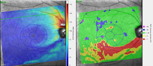
Example of the new Thickness Map and Thickness Deviation Map with temporal inferior arcuate RNFL defect.Hemel Hempstead, United Kingdom – SPECTRALIS Glaucoma Module Premium Edition (GMPE) customers having installed the SPECTRALIS release 6.12.1 can now benefit from the discriminative power of the macular reference database with the addition of thickness deviation maps for retina, retinal nerve fibre, ganglion cell and inner plexiform layers. This feature is free of charge to all SPECTRALIS owners with GMPE Module. The Deviation Maps update offers:
Thickness deviation maps, which highlight whether the thickness measurements of each retinal layer are within or outside of normal limits in comparison to a reference database, making the regions and extent of structural thickening or thinning clearly discernible.
New thickness maps with a perceptually uniform colour scale that make changes in retinal thickness more easily perceived by the human eye in comparison to the traditional hue colour scale.
Classification chart with values derived from a Garway-Heath 6 sector grid, accurately anatomically aligned on the macular according to the individual anatomy of each eye using the Anatomic Positioning System (APS).
“The comparison of thickness measurements with the reference database enhances the diagnostic value of thickness maps and improves the visualisation of structural defects consistent with glaucoma”, explains Tim Cole, Clinical Market Development Manager, Heidelberg Engineering UK. “Heidelberg Engineering is well renowned for having revolutionised glaucoma diagnostics after bursting onto the scene with the Heidelberg Retina Tomograph (HRT) in 1999. The launch of Deviation Maps, and the Glaucoma Hood Report earlier in 2019, shows a continued commitment by the company to developing innovative and clinically validated diagnostic solutions for glaucoma.”
Contact Heidelberg Engineering to find out how to enable Deviation Maps functionality or to enquire about upgrading your SPECTRALIS with Glaucoma Module Premium Edition by calling 01442 502 330 or emailing Info-UK@HeidelbergEngineering.com.
MP Sir Mike Penning visits Heidelberg Engineering to promote World Glaucoma Week
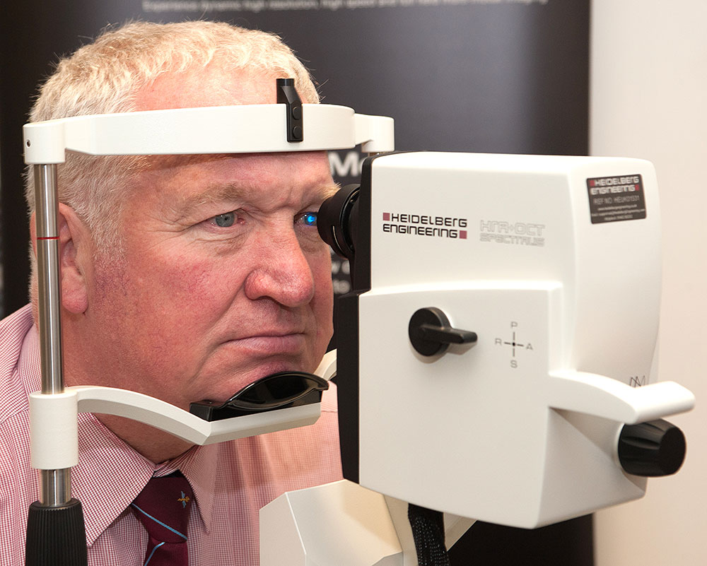
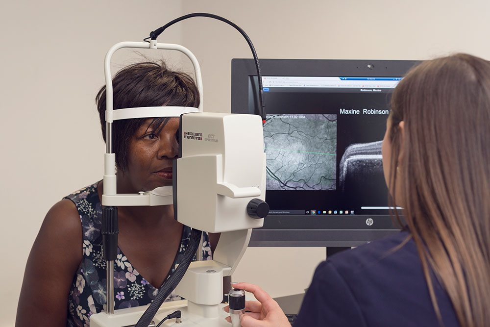
MP Sir Mike Penning sitting in front of a SPECTRALISHemel Hemstead, UK – Sir Mike Penning MP marked the start of World Glaucoma Week (11-17 March) when he called into his Hemel Hempstead constituency company Heidelberg Engineering which provides scanning technology for advanced eye health check.
Sir Mike had his eyes examined on the SPECTRALIS OCT (Optical Coherence Tomographer) which can reveal the first signs of eye disease years before they might become apparent to the patient. He was delighted to learn that he showed no signs of developing glaucoma – a leading cause of preventable blindness.
Krysten Williams, UK Director of Heidelberg Engineering explained –
“Sir Mike has highlighted the need for a regular eye health check as glaucoma – has no obvious signs in the early stages of the disease. As pressure builds in the eye, the optic nerve can be irreversibly damaged, but the patient is not normally aware of this happening. The patient may not even notice vision loss because the brain cleverly compensates for any missing information. This is why regular screening is so important.”
Sir Mike toured the company’s training centre, met staff and learned about the company’s mission to support a sustainable healthcare model that devolves early detection and monitoring of eye disease into the community. This would facilitate treatment at the right time to combat avoidable sight loss.
“As Sir Mike has a keen interest in healthcare, it was a great opportunity to discuss the effects of vision loss and the tremendous opportunities to use the latest image management technologies to reduce the growing burden of an ageing population on the health service with distributed care models,” added Krysten.
Sir Mike said –
“It was fascinating to visit Heidelberg Engineering and learn about how their advanced scanning technology is helping prevent blindness through identification of patients with the early stages of glaucoma. I would encourage everyone to have regular eye checks whether they think they need new glasses or not!”
Professor John Nolan, BSc, PhDHemel Hemstead, UK – The significance of macular nutrition for good eye health comes into sharp focus at Optrafair with a CET lecture from Professor John Nolan, BSc, PhD, who leads international research in this field.
Sponsored by Heidelberg Engineering, the insight into current understanding of macular pigment significance, takes place on Sunday 15th April at the NEC, Birmingham show. Places are booking up fast so registering is recommended via www.optrafair.co.uk/education/cet-theatre
Following several years of research, Professor Nolan will share his internationally respected findings of how the macular pigment influences ocular health and cognitive function. Guidance on how to interpret clinical trial data and the impact on visual and cognitive performance will be covered, as well as how assessing macular nutrition could motivate patients to consider positive lifestyles changes.
Christopher Mody, Heidelberg Engineering Professional Services Manager, explained –
“There is a lot of clinical noise about macular nutrition, but we are bringing to Optrafair a world class authority on the subject who will provide much needed clarity. Professor Nolan will summarise the novel protocol for using dual wavelength auto-fluorescence for assessing macular nutrition. Using data from the European Research Council funded CREST clinical trials he will make recommendations on using this knowledge to take a more holistic approach to patient care.”
Heidelberg Engineering will have a strong presence at Optrafair and its team of experts will be on hand to discuss all aspects of monitoring eye health from High Street optometry practices.
Tim Cole joins the Heidelberg Engineering Academy Team
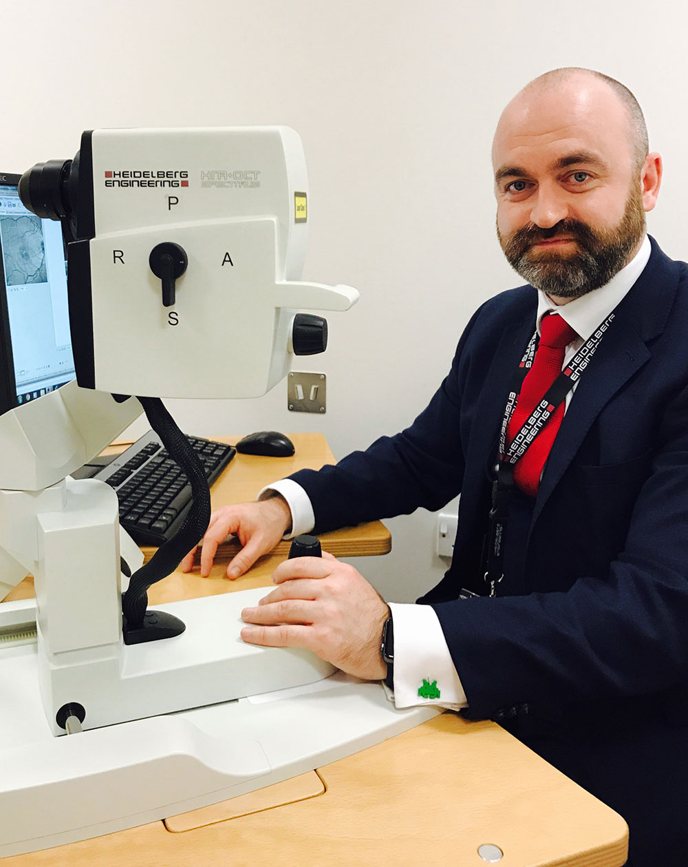
Tim Cole, Heidelberg Engineering AcademyHemel Hempstead, UK – Heidelberg Engineering has recruited imaging expert Tim Cole – known to many in the optical profession for his OCT knowledge.
Following six years with Topcon, Tim joins Heidelberg Engineering as Clinical Market Development Manager and will be supporting Christopher Mody, Director of Clinical Affairs , in educating clinicians and supporting their research projects.
Tim, who trained as an NHS Ophthalmology photographer at Manchester Royal Eye Hospital, is highly impressed with the Heidelberg Engineering technology –
“The SPECTRALIS is amazing. I always knew it to be an exceptional piece of technology and the more I have learned about its functionality the more amazed I am. The precision of SPECTRALIS in capturing confocal scanning laser images of the retina surpasses anything that I have ever seen before.”
Tim, who is well known in both the High Street optometry world and the eye hospital community, has been warmly welcomed by the Heidelberg Engineering team, as UK Director Krysten Williams, said:
“We are very focused on strengthening our Academy team to hold our position as leaders in clinical education. Tim comes to us with quite a pedigree and will help us with education at every level from research, to clinic, and High Street optometry.”
To learn more about the Heidelberg Engineering Academy please contact: 01442 502 330
Specsavers and Heidelberg Engineering set to transform Community Eyecare in the UK
Hemel Hempstead, UK – Heidelberg Engineering has designed the SPECTRALIS SPIRIT, a bespoke OCT imaging platform – which achieves high definition images in just three clicks – to support Specsavers’ ambition to offer patients a choice of the best possible technology in their eye examination by installing an OCT in every practice.
The new SPECTRALIS SPIRIT is set to transform eyecare in the community at a time when hospital services are increasingly stretched. Providing excellent image quality, accuracy and reproducibility, it measures up to one micron of change over time – which sets the technology apart from competitors. With an emphasis on glaucoma diagnosis and care, the SPECTRALIS SPIRIT uses highly sensitive and specific parameters for early detection of glaucoma, minimising false positive referrals and providing the clinician with an easy-to-interpret traffic light signal report.
To understand how the Specsavers’ partnership integrates with the existing SPECTRALIS business, Krysten Williams, Director of Heidelberg Engineering UK, stressed the differentiation of the two models and the roles they play –
“In past years the challenge was to find and design an imaging solution to meet the demand for a high-volume retail environment, and to ensure that both optometrists and ophthalmologists can embrace shared care with confidence,” said Krysten Williams.
“This strategic move is a highly significant contribution to advancing eyecare in the community. Those who have already invested in the SPECTRALIS platform can be confident that the full range of functionality and imaging modalities of the retinal specialists’ OCT of choice, as well as their own expertise continues to set them apart.”
“We have just one ophthalmologist per 50,000 people in the UK. By comparison, in Germany the figure is one per 11,100 people. High Street optometry could help to relieve the burden on hospital eye departments, who are faced with an ageing demographic, and an alarming rise in age-related eye disease. For over 6 years Heidelberg Engineering UK has been dedicated to providing high level clinical education to optometrists and we have seen those who invested in technology and their own education raise the standard of patient care in their community.“
“The collaboration with Specsavers raises the bar for diagnostic technology and education in all Specsavers practices and, in turn, encourages the NHS to recognise the profession as the logical choice for commissioning and paying for services provided in the community. The business opportunity for diagnosis and care is here for the entire profession to seize. The SPECTRALIS SPIRIT is available to all optometrists, allowing them to build their clinical confidence by starting with the essentials of OCT and upgrading to additional imaging modalities that enhance their clinical decision making as their practice grow.”
Paul Morris, Specsavers’ Optometrist Director of Professional Advancement, spoke of the enthusiasm of Specsavers ‘practices to roll out OCT, and the company’s “significant” investment in training –
“OCT is the future of optometry. Our partners have shown a passion for the opportunity to offer the latest in imaging technology to better identify and manage the long-term eye health of patients. Our relationship with Heidelberg Engineering is new, but the quality of their OCT devices needs no explanation as the company is synonymous with leading-edge technology. I am confident that we will have a long and mutually beneficial relationship.”
If you have questions about Heidelberg Engineering’s partnership with Specsavers or about the SPECTRALIS SPIRIT please call 01442 502 330 or email Info-UK@HeidelbergEngineering.com
New study proves advantages of MultiColor imaging over fundus photography for early diagnosis and monitoring of AMD
Heidelberg, Germany – A research group from the Centre for Public Health at the Queen’s University in Belfast, Northern Ireland, has just published the results of a study comparing SPECTRALIS® MultiColor imaging and traditional color fundus photography for the detection of features of early and late age-related macular degeneration (AMD). The study’s findings demonstrate the benefits of SPECTRALIS MultiColor imaging for increased sensitivity and specificity.
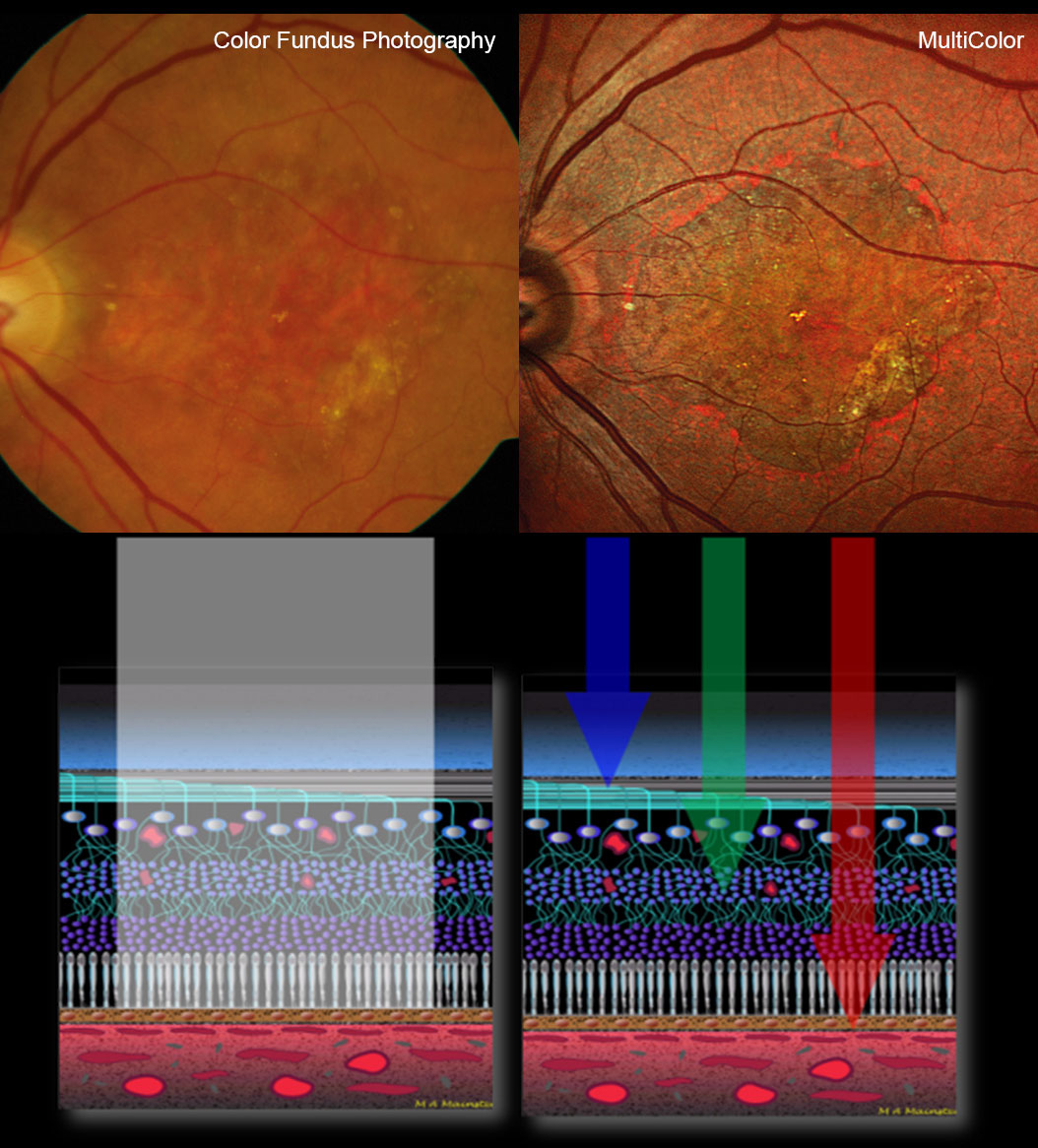
The SPECTRALIS MultiColor Module is an imaging modality which utilizes confocal scanning laser technology instead of white light to visualize the retina.The study, which was published digitally in August and will appear in the November issue of Retina, is titled: “Identifying features of early and late AMD: A comparison of MultiColor versus traditional color fundus photography”. The authors are Katie W. Graham, Usha Chakravarthy, Ruth E. Hogg, K. Alyson Muldrew, Ian S. Young and Frank Kee, all of the Centre for Public Health, Queen’s University in Belfast, Northern Ireland.
The goal of the study was to compare the MultiColor Module of the SPECTRALIS diagnostic imaging platform to color fundus photography (CFP), which is currently considered the gold standard examination for the recognition and classification of features of early and late AMD. The SPECTRALIS MultiColor Module, on the other hand, is a newer imaging modality which utilizes confocal scanning laser technology with light of discrete wavelengths instead of standard optics and white light to visualize the retina.
MultiColor was proven to have higher sensitivity than CFP for the detection of early AMD features using a sample of 105 eyes. In cases with discrepancies, an analysis of OCT also showed better agreement with MultiColor for all AMD lesions, with the exception of hemorrhage and non-geographic atrophy hypopigmentation. For pigment clumping, CFP and MultiColor were in equal agreement to OCT.
MultiColor imaging was able to identify soft drusen in 85%, reticular drusen in 83%, and atrophy and fibrosis in 100% of cases where these abnormalities were seen on CFP. However, when using MultiColor as the basis for analysis, CFP was less sensitive. In this analysis, soft drusen were identified in only 58%, reticular drusen in 28%, atrophy in 83%, and fibrosis in 68% of the cases where such changes were seen on MultiColor images.
“This systematic analysis of color fundus photography versus MultiColor demonstrated the robustness of MultiColor imaging in the detection of early AMD features. The ability to delineate atrophy and fibrosis in late stage AMD gives added value in the clinical setting, as these components of the wet AMD lesion have an important impact on visual function”, said Usha Chakravarthy, Professor of Ophthalmology and Vision Sciences at Queen’s University, Belfast, Northern Ireland.
Other clinicians also familiar with the MultiColor Module appreciate its advantages: “With the advent of MultiColor imaging we can obtain high-resolution fundus images for a precise study of the morphometric changes of the retina and combine them with structural OCT scans of up to 55 degrees. This gives us the possibility to monitor changes from visit to visit in both imaging modalities. In addition, at our clinic we can even obtain an OCTA over a MultiColor image, selecting the area of particular interest. And all of that, without having the patient moving from one system to another”, said Roberto Gallego-Pinazo, MD, Retina Specialist of the Macula Unit at Oftalvist in Valencia, Spain.
See structure at different depths
MultiColor is able to achieve such sensitivity and specificity due to its confocal scanning laser imaging technology, which uses three laser wavelengths (blue, green and infrared) simultaneously to provide diagnostic images that show distinct structures at different depths within the retina. As a result, the high-resolution MultiColor images can be used to detect and delineate structures and pathologies not visible on ophthalmoscopy and fundus photography.
MultiColor images can and should ideally be visualized both as a composite image combining all three wavelengths and also as individual blue, green and infrared reflectance images. Each of the three reflectance images offers unique details due to the penetration depth and reflectance properties of each individual wavelength used. The infrared reflectance image shows deeper structures in the choroid and the retinal pigment epithelium. The green reflectance image is useful for examining blood, blood vessels, and exudates. The blue reflectance image is best for identifying changes in superficial retinal structures, like epiretinal membranes or retinal nerve fiber layer defects.
MultiColor as part of a multimodal approach
“It is encouraging to see the clinical significance of the MultiColor Module proven in a large study related to a prevalent pathology such as AMD. Beyond the clinical value, clinicians also appreciate the fact that MultiColor images can be acquired through undilated pupils and in patients with media opacity or even nystagmus. While we don’t see MultiColor as a substitute for color fundus photography, which remains useful for documentation purposes, it is a particularly useful diagnostic tool that can be combined with other imaging modalities”, said Dr. Kester Nahen, Managing Director at Heidelberg Engineering.
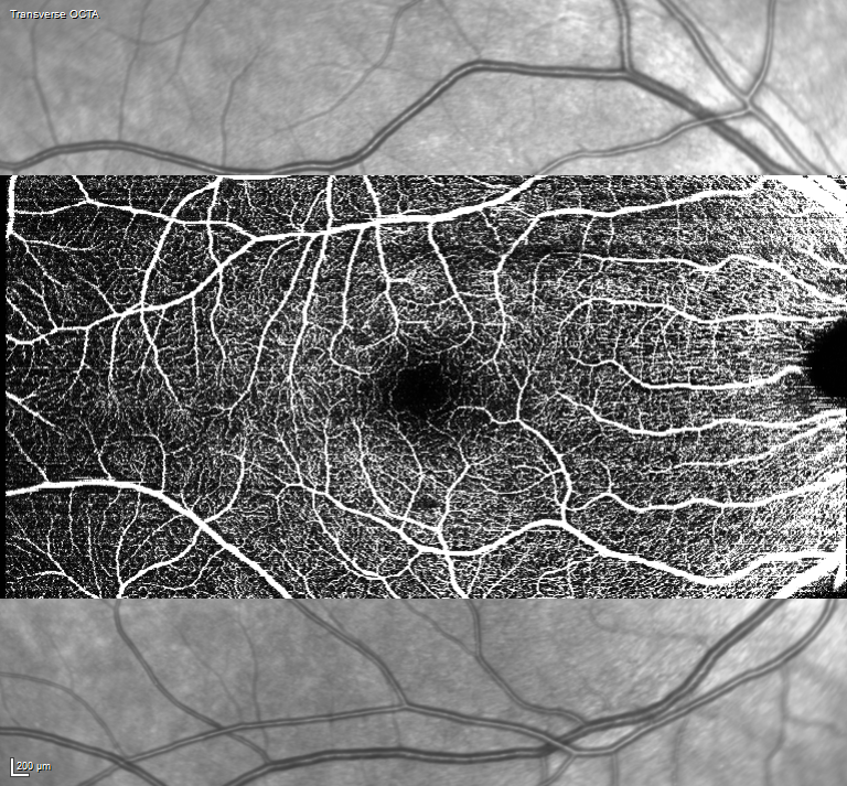
SPECTRALIS OCT Angiography Module update enhances speed and expands the field of view
Hemel Hempstead, UK – Heidelberg Engineering has released an update for the SPECTRALIS OCT Angiography Module, which is now available to all new and existing customers. New benefits include a significantly faster image processing time and the ability to customise the field of view of the OCTA image to suit the level of detail required for clinical assessment. The field of view ranges from a high resolution 10°x10° at 5.7µm/pixel lateral resolution, which is suitable for indicating the presence or absence of flow in even the smallest capillaries, to a full field view of 30°x15° at 11µm/pixel.
“The unique algorithm utilised in the OCT Angiography Module for SPECTRALIS separates vascular flow from structure using a sophisticated statistical analysis that, put simply, assigns flow as white and structure as black in the image” explains Christopher Mody, Director of Clinical Services. “This approach, combined with patented TruTrack Active Eye Tracking, ensures there are no grey areas of uncertainty in the OCTA image and produces the highest resolution OCTA images in any field of view.”
Also included in the update is the fusion of high resolution OCT images with flow. The flow is colour-coded as yellow on the OCT image to allow the clinician to visualise its precise location in abnormal vessels to aid assessment of pathology.
Flex Module for the SPECTRALIS used in first human aqueous angiography study
SPECTRALIS Flex ModuleHemel Hempstead, UK – A pilot study, headed by Dr Alex Huang, Doheny Eye Institute, USA, has confirmed the efficacy of live aqueous angiography on human patients in need of cataract surgery. Using the Flex Module for SPECTRALIS, the study demonstrated the ability to safely perform such a procedure, allowing for refinement of the surgical techniques needed in order to develop a concrete protocol for the next phase of the trial. The results of this study further confirmed the outcomes of previous ex-vivo and in-vivo non-human primate studies.
SPECTRALIS Flex Module, with its movable stand and an adjustable arm, enables all SPECTRALIS examinations, using any imaging modality, to be performed on patients lying down in the supine position. “Minimally invasive trabecular bypass/ablation glaucoma surgeries (MIGS) have shown intraocular pressure reduction, but the magnitude is variable and unpredictable” explains Christopher Mody, Director of Clinical Services. “Now that we can examine individual patients with the SPECTRALIS Flex Module to understand the physiology of aqueous humor outflow pathways, it may be possible to enhance the magnitude of intraocular pressure lowering and its predictability.”
The next phases of Aqueous Angiography research will aim to determine outflow differences between glaucomatous eyes and those without glaucoma. Dr. Huang and his research team also intend to show whether Aqueous Angiography-guided MIGS placement is feasible and whether such a procedure reveals improvement in outflow pathways and potential enhancement in efficacy of MIGS.
The Flex Module will be available for delivery in June 2017. Find out more about other applications for the new Flex Module for the SPECTRALIS Imaging Platform by emailing info-UK@HeidelbergEngineering.com.