Heidelberg, Germany – A research group from the Centre for Public Health at the Queen’s University in Belfast, Northern Ireland, has just published the results of a study comparing SPECTRALIS® MultiColor imaging and traditional color fundus photography for the detection of features of early and late age-related macular degeneration (AMD). The study’s findings demonstrate the benefits of SPECTRALIS MultiColor imaging for increased sensitivity and specificity.
The goal of the study was to compare the MultiColor Module of the SPECTRALIS diagnostic imaging platform to color fundus photography (CFP), which is currently considered the gold standard examination for the recognition and classification of features of early and late AMD. The SPECTRALIS MultiColor Module, on the other hand, is a newer imaging modality which utilizes confocal scanning laser technology with light of discrete wavelengths instead of standard optics and white light to visualize the retina.
MultiColor was proven to have higher sensitivity than CFP for the detection of early AMD features using a sample of 105 eyes. In cases with discrepancies, an analysis of OCT also showed better agreement with MultiColor for all AMD lesions, with the exception of hemorrhage and non-geographic atrophy hypopigmentation. For pigment clumping, CFP and MultiColor were in equal agreement to OCT.
MultiColor imaging was able to identify soft drusen in 85%, reticular drusen in 83%, and atrophy and fibrosis in 100% of cases where these abnormalities were seen on CFP. However, when using MultiColor as the basis for analysis, CFP was less sensitive. In this analysis, soft drusen were identified in only 58%, reticular drusen in 28%, atrophy in 83%, and fibrosis in 68% of the cases where such changes were seen on MultiColor images.
“This systematic analysis of color fundus photography versus MultiColor demonstrated the robustness of MultiColor imaging in the detection of early AMD features. The ability to delineate atrophy and fibrosis in late stage AMD gives added value in the clinical setting, as these components of the wet AMD lesion have an important impact on visual function”, said Usha Chakravarthy, Professor of Ophthalmology and Vision Sciences at Queen’s University, Belfast, Northern Ireland.
Other clinicians also familiar with the MultiColor Module appreciate its advantages: “With the advent of MultiColor imaging we can obtain high-resolution fundus images for a precise study of the morphometric changes of the retina and combine them with structural OCT scans of up to 55 degrees. This gives us the possibility to monitor changes from visit to visit in both imaging modalities. In addition, at our clinic we can even obtain an OCTA over a MultiColor image, selecting the area of particular interest. And all of that, without having the patient moving from one system to another”, said Roberto Gallego-Pinazo, MD, Retina Specialist of the Macula Unit at Oftalvist in Valencia, Spain.
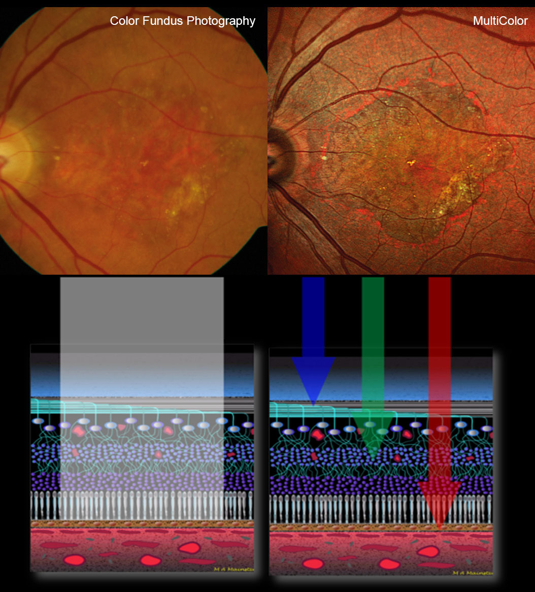
See structure at different depths
MultiColor is able to achieve such sensitivity and specificity due to its confocal scanning laser imaging technology, which uses three laser wavelengths (blue, green and infrared) simultaneously to provide diagnostic images that show distinct structures at different depths within the retina. As a result, the high-resolution MultiColor images can be used to detect and delineate structures and pathologies not visible on ophthalmoscopy and fundus photography.
MultiColor images can and should ideally be visualized both as a composite image combining all three wavelengths and also as individual blue, green and infrared reflectance images. Each of the three reflectance images offers unique details due to the penetration depth and reflectance properties of each individual wavelength used. The infrared reflectance image shows deeper structures in the choroid and the retinal pigment epithelium. The green reflectance image is useful for examining blood, blood vessels, and exudates. The blue reflectance image is best for identifying changes in superficial retinal structures, like epiretinal membranes or retinal nerve fiber layer defects.
MultiColor as part of a multimodal approach
“It is encouraging to see the clinical significance of the MultiColor Module proven in a large study related to a prevalent pathology such as AMD. Beyond the clinical value, clinicians also appreciate the fact that MultiColor images can be acquired through undilated pupils and in patients with media opacity or even nystagmus. While we don’t see MultiColor as a substitute for color fundus photography, which remains useful for documentation purposes, it is a particularly useful diagnostic tool that can be combined with other imaging modalities”, said Dr. Kester Nahen, Managing Director at Heidelberg Engineering.
Journal Article Review (Business Lounge login required)


